Does A Head Ct Scan Help Diagnose Multiple Sclerosis If A Ct Scan Is Normal Is It Still Likely That Multiple Sclerosis Can Be Present
Dr. Oscar DellingerDr. Duane GelsMultipleDr. Bennett Machanic
Ask U.S. doctors your own question and get educational, text answers — it’s anonymous and free!
Ask U.S. doctors your own question and get educational, text answers — it’s anonymous and free!
HealthTap doctors are based in the U.S., board certified, and available by text or video.
Parkinsons Is Easily Identified By A Pathologist But Is Difficult To Diagnose For A Clinician
When I state that Parkinson’s Disease is “well defined,” I mean that the pathologist, looking at slides of the brain under a microscope, can say, knowing nothing about the patient, that the person had PD. Parkinson’s Disease causes certain, easy to detect changes that are seen under the microscope. This, of course, requires that the person is dead, which isn’t much use to that particular person or his family. The absence of a reliable test, in life, means then that there is room for mistakes. And, in fact, we make a fair number of mistakes, and I’ll discuss below the types of cases in which we are most likely to make mistakes. Unfortunately, we are never in the position of being 100% certain that we’ve diagnosed someone correctly, until the autopsy.
Should I Get A Datscan Or Pet Scan To Confirm My Diagnosis Of Parkinsons Disease
You can find out more about NPF’s National Medical Director, Dr. Michael S. Okun, by also visiting the NPF Center of Excellence, University of Florida Center for Movement Disorders & Neurorestoration.
This past month, the FDA approved DaTscan , a radiopharmaceutical agent which is injected into a patient’s veins in a procedure referred to as SPECT imaging. DaTscan is an important addition because it is anticipated to be more widely available than other techniques and it has received several major endorsements from leading scientists.
One of the most frequently asked questions about Parkinson’s disease on NPF’s “Ask the Doctor” web-based forum is whether or not to pursue DaT or PET scanning to confirm a diagnosis of Parkinson’s disease. In this month’s What’s Hot column, we offer a review of the subject in light of the recent FDA approval.
The new DaT scans use a substance that “tags” a part of a neuron in the brain where dopamine attaches to it, showing the density of healthy dopamine neurons. Thus, the more of the picture that “lights up”, the more surviving brain cells. If the parts of the brain where dopamine cells should be remain dark in the scan, an expert reader may diagnose early brain degeneration. This could mean either Parkinson’s disease or parkinsonism.
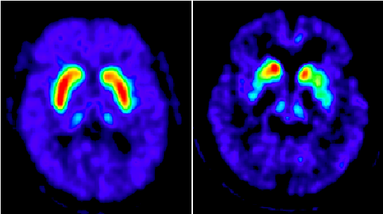
An example DaTscan is shown below and it demonstrates essential tremor on the left , and a parkinsonian syndrome on the right .
Imaging Studies Can Differentiate Parkinsons From Other Causes Of Parkinsonism
Catherine L. Gallagher, MD
Although Parkinson’s disease remains a clinical diagnosis, imaging studies are an important ancillary test for differential diagnosis of movement disorders. Imaging studies may be used to rule out structural and other causes of parkinsonian symptoms. Single-photon emission computed tomography scans using labeled tracers for dopamine transporters can also be used to confirm parkinsonism or differentiate PD from secondary causes of parkinsonian motor symptoms. Finally, imaging studies are being used in research to better understand the pathophysiology of PD and elucidate causative mechanisms that could be therapeutic targets in the future.
Diagnosis Of Parkinsons Via Datscan And Clinical Exam Are Similarly Accurate
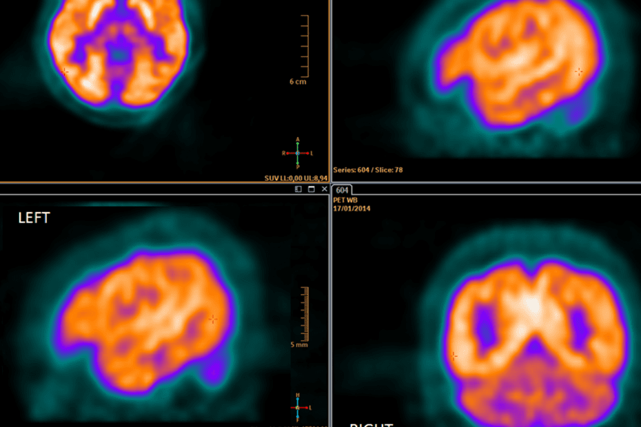
Despite the DaTscan being available to help diagnose Parkinson’s, in most clinical situations, a DaTscan will not add information to what can be gleaned from the clinical exam. One study actually demonstrated that the accuracy of diagnosis in early PD was the same whether the diagnosis was reached using clinical exam or using DaTscan.
What’s Hot In Pd An Update On Dat Scanning For Parkinsons Disease Diagnosis
In 2011, the FDA approved a diagnostic test for Parkinson’s disease. The DaTscan is a radiopharmaceutical agent which is injected into a patient’s veins in a procedure referred to as SPECT imaging. DaTscan, when it was approved, was considered an important addition to the armamentarium of the bedside clinician. In 2011 I wrote a What’s Hot column on DAT scanning, and this month I will update that posting and bring everyone up to date on the impact of this test.
One of the most frequently asked questions about Parkinson’s disease on NPF’s “Ask the Doctor” web-based forum is whether or not to pursue DaT or PET scan to confirm a diagnosis of Parkinson’s disease. The short answer is that the DaT test is over-used in clinical practice, and is only FDA approved to distinguish potential Parkinson’s disease from essential tremor. In fact, the test only tells the clinician if there is an abnormality in the dopamine transporter, and does not actually diagnose Parkinson’s disease . PET is also overused, though it can be a more powerful diagnostic tool when in the right expert hands.
The new DaT scans use a substance that “tags” a part of a neuron in the brain where dopamine attaches to it, thus showing the density of healthy dopamine neurons. Thus, the more of the picture that “lights up”, the more surviving brain cells. Dark areas could mean either Parkinson’s disease or parkinsonism.
Selected References
What Is Essential Tremor And How Is It Different To A Parkinsons Tremor
A tremor is a rhythmical, involuntary movement that affects a part of the body, such as the hand.
Essential tremor is the most common type of tremor. It’s most noticeable when your hands are doing something and it usually affects both the right and left sides of the body equally. Essential tremors often lessen when your body is resting.
Unlike an essential tremor, a Parkinson’s tremor is most obvious when the affected body part is resting and tends to be less noticeable with movement. It usually starts on one side of the body and may progress to the other side as Parkinson’s develops.
The time it takes to get a diagnosis can vary from person to person. Some people may receive a diagnosis of Parkinson’s quite quickly, but for others it may be a long process. This can be due to a number of things, including your medical history, your age and what symptoms you have.
Your specialist may wish to rule out other causes of your symptoms first and see how you respond to treatment. This may take some time, and, as already mentioned, there is currently no definitive test for Parkinson’s.
How you respond to treatment may help your specialist make a diagnosis. Keeping a diary or record of your symptoms will give the specialist more information to guide their decision.
Because the symptoms of Parkinson’s are sometimes similar to other forms of parkinsonism, people can sometimes be misdiagnosed.
Differences Between Parkinsons Disease And Atypical Parkinsonism
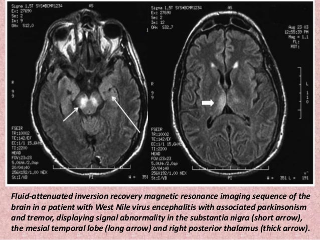
The symptoms of Parkinson’s disease and atypical parkinsonism overlap, and in a clinical setting, it can be hard to tell if a patient has one or the other. Atypical parkinsonism is diseases that present some of the signs and symptoms of Parkinson’s Disease but do not respond well to drug treatment. With an MRI, your doctor can help to make the diagnosis more accurate, which is essential for quality treatment. Additionally, an MRI can also help your medical team to determine if you have a certain type of atypical parkinsonism. This can help to create a prognosis and guide your treatment options.
Determining Diagnosis Through Response To Parkinsons Medication
If a person’s symptoms and neurologic examination are only suggestive of Parkinson’s disease or if the diagnosis is otherwise in doubt, the physician may, nevertheless, prescribe a medication intended for Parkinson’s disease to provide additional information. In the case of idiopathic Parkinson’s, there is typically a positive, predictable response to Parkinson’s disease medication; in the case of some related Parkinsonian syndromes, the response to medication may not be particularly robust, or it may be absent entirely.
Unfortunately, there are no standard biological tests for the disease, such as a blood test. However, researchers are actively trying to find biomarkers in blood and other bodily fluids that could help confirm the diagnosis.
Brain Imaging And Other Tools To Aid Diagnosis Of Parkinsons
In addition to taking a history and performing a detailed neurologic examination, physicians sometimes use brain imaging to help support a particular diagnosis. However, these studies have their limitations in the diagnosis of Parkinson’s disease and are typically used only in select patients. Brain imaging is not routinely performed by neurologists or movement disorder specialists when they are considering a diagnosis, especially if the person’s symptoms strongly suggest to the physician that idiopathic Parkinson’s disease is the correct diagnosis.
Helping diagnose Parkinson’s with DaTscan and other tests
Rather, use of imaging is most helpful when the diagnosis is uncertain, or when physicians are looking for changes in the brain that are more typical of one of several Parkinsonian syndromes and other conditions that can mimic Parkinson’s. Imaging studies to evaluate Parkinson’s disease and Parkinsonian syndromes include magnetic resonance imaging , which examines the structure of the brain, and DaTscan, an imaging test approved by the Food and Drug Administration to detect the dopamine function in the brain. A DaTscan may help differentiate idiopathic Parkinson’s disease from certain other neurologic disorders. Most physicians’ offices will have access to MRI; however, DaTscan imaging may only be available at larger hospitals or medical centers.
Accurate Parkinson’s Diagnosis & Other Movement Disorders movement disorder neurologist
Making an accurate diagnosis in the early stages of Parkinson’s disease and other movement disorders can be difficult. The beginning signs and symptoms may be similar to those related to many other conditions or to the effects of normal aging. For this reason, observation of the patient may be required for some time until the symptoms are consistently present.
Datscan: A Test To Help In The Diagnosis Of Parkinsons
In 2011, the Food and Drug Administration approved an imaging test to help diagnose PD. In this test, a radioactive tracer, Ioflupane 123I, also known as DaTscan, is injected into the blood, where it circulates around the body and makes its way into the brain. It attaches itself to the dopamine transporter, a molecule found on dopamine neurons. Several hours after the tracer has been injected, special imaging equipment scans the head to detect the presence of DaTscan.
People with PD will typically have a smaller signal in a part of the brain called the striatum, where the ends of the dopamine neurons are meant to be. Here is a normal scan on the left, which would indicate a healthy dopamine system, next to an abnormal scan on the right, which would indicate an unhealthy dopamine system.
It is important to note that conventional MRI imaging will appear normal in PD and is therefore not helpful in confirming the diagnosis. Other atypical parkinsonian conditions, such as vascular parkinsonism however, can have abnormalities on MRI, so the test may be done to rule out other diagnoses.
Mri Brain Scans Detect People With Early Parkinson’s
Oxford University researchers have developed a simple and quick MRI technique that offers promise for early diagnosis of Parkinson’s disease.
The new MRI approach can detect people who have early-stage Parkinson’s disease with 85% accuracy, according to research published in Neurology, the medical journal of the American Academy of Neurology.
‘At the moment we have no way to predict who is at risk of Parkinson’s disease in the vast majority of cases,’ says Dr Clare Mackay of the Department of Psychiatry at Oxford University, one of the joint lead researchers. ‘We are excited that this MRI technique might prove to be a good marker for the earliest signs of Parkinson’s. The results are very promising.’
Claire Bale, research communications manager at Parkinson’s UK, which funded the work, explains: ‘This new research takes us one step closer to diagnosing Parkinson’s at a much earlier stage – one of the biggest challenges facing research into the condition. By using a new, simple scanning technique the team at Oxford University have been able to study levels of activity in the brain which may suggest that Parkinson’s is present. One person every hour is diagnosed with Parkinson’s in the UK, and we hope that the researchers are able to continue to refine their test so that it can one day be part of clinical practice.’
We think that our MRI test will be relevant for diagnosis of Parkinson’s
Dr Michele Hu
Fact Sheet: Molecular Imaging And Parkinsons Disease
Parkinson’s disease is a brain disorder that leads to motor symptoms, such as shaking and difficulty with walking, movement, and coordination. Patients with PD may also experience non-motor symptoms, such as changes in mood or cognition. PD is a progressive and neurodegenerative disorder.
The disease most often develops after age 50 and symptoms vary from patient to patient.
PD is the most common among a group of movement disorders called Parkinsonian syndromes, all of which result from a loss of dopamine-producing neurons and have similar symptoms. PD is the second most common neurodegenerative disorder after Alzheimer’s disease.
- Nearly one million will be living with Parkinson’s disease in the U.S. by 2020, which is more than the combined number of people diagnosed with multiple sclerosis, muscular dystrophy and Lou Gehrig’s disease
- Approximately 60,000 Americans are diagnosed with PD each year.
- More than 10 million people worldwide are living with PD.
- Incidence of Parkinson’s disease increases with age, but an estimated four percent of people with PD are diagnosed before age 50.
- Men are 1.5 times more likely to have Parkinson’s disease than women.
What is molecular imaging?
Molecular imaging is a type of medical imaging that provides unique, detailed pictures of what is happening inside the brain at the molecular and cellular level. Other diagnostic imaging procedures—such as x-rays, computed tomography and ultrasound—predominantly offer anatomical pictures.
What Doctors Look For When Diagnosing Parkinsons
Certain physical signs and symptoms — noticed by the patient or his or her loved ones — are usually what prompt a person to see the doctor. These are the symptoms most often noticed by patients or their families:
-
Shaking or tremor: Called resting tremor, a trembling of a hand or foot that happens when the patient is at rest and typically stops when he or she is active or moving
-
Bradykinesia: Slowness of movement in the limbs, face, walking or overall body
-
Rigidity: Stiffness in the arms, legs or trunk
-
Posture instability: Trouble with balance and possible falls
Once the patient is at the doctor’s office, the physician:
-
Takes a medical history and does a physical examination.
-
Asks about current and past medications. Some medications may cause symptoms that mimic Parkinson’s disease.
-
Performs a neurological examination, testing agility, muscle tone, gait and balance.
How A Head Ct Scan Can Detect Alzheimers Disease
A head CT scan looks at the structure of your brain. This scan can detect issues such as tumors, hemorrhages, and strokes, which can all mimic the symptoms of Alzheimer’s, but in addition to helping you rule out those conditions, a CT scan can also detect the loss of brain mass that’s associated with Alzheimer’s disease.
Other Tests Used To Diagnose Parkinsons Disease
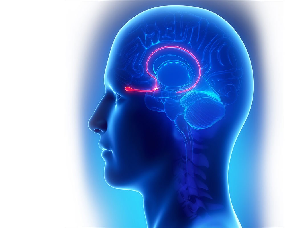
Diagnosis of PD is generally made using a medical history and a physical exam. In addition to these tests, the patient presenting with Parkinsonian symptoms is usually given treatment with the medication levodopa. The relief of symptoms with levodopa is a sign that symptoms are caused by PD.6
An active area of research is discovering “biomarkers”, which are molecules in the blood, urine, or cerebrospinal fluid that can reliably diagnose Parkinson’s disease.
There Is No Test To Diagnose Parkinsons Disease
Although Parkinson’s disease is a specific, well defined disease that can be reliably diagnosed at autopsy, it is defined in life by clinical criteria. This means that the diagnosis rests entirely on the information that the patient provides plus the physical examination. There is no test to diagnose PD. When a doctor orders a test when evaluating a patient who he thinks has PD, the test is really being obtained to make sure that there isn’t some other disorder that might look like PD. When I am convinced that a patient has Parkinson’s Disease I order no tests at all. No CAT scan, no MRI, no EEG, no spinal tap, no blood tests. Nothing. The reason is that if any of these tests was to show an abnormality, it would mean to me that the patient had Parkinson’s Disease plus some other disorder that has nothing to do with why the patient was seeing me in the first place. Generally I’d rather not know.
Why Is A Pet Scan Used In Parkinson’s Disease
For patients with Parkinson’s disease , a PET scan is used to assess activity and function of brain regions involved in movement. However, doctors may request a PET scan for many different reasons. Aside from potential problems in the brain and spinal cord, the test can also be used to diagnose heart problems as well as certain kinds of cancer, including breast, brain, lung, colon, and prostate cancers and lymphoma.
Why Choose Independent Imaging For My Datscan
Independent Imaging has been putting patients first for over 30 years. Our imaging centers in Wellington, Belle Glade, Lake Worth, and Royal Palm Beach, Florida are fully accredited by the American College of Radiology. Our board-certified radiologists are happy to answer any questions you may have, and we offer extended hours six days a week to accommodate our patients and referring physicians.
If you or a loved one would like to schedule a DaTSCAN™, or any of the other imaging services we offer, please call 795-5558 today, or submit an appointment request online.
A Brain Scan That Could Diagnose Parkinson’s
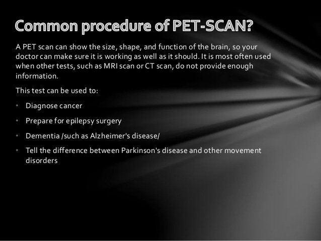
New research, published in the scientific journal Radiology, highlights the potential of using a type of brain scan as a diagnostic tool in Parkinson’s.
The Nottingham University Hospital researchers, funded by Parkinson’s UK, used an MRI brain scan to study changes in a pigment in the part of the brain affected by Parkinson’s.
In 69 participants, they found that the brain scan was highly accurate at identifying those with mild or moderate Parkinson’s – highlighting the potential of this technique as a diagnostic test.
What Can I Expect On The Day Of The Ct Scan
Please allow at least one hour for your CT scan. Most scans take from 15 to 60 minutes.
Depending on the type of scan you need, a contrast material may be injected intravenously so the radiologist can see the body structures on the CT image.
After the contrast agent is injected, you may feel flushed, or you may have a metallic taste in your mouth. These are common reactions. If you experience shortness of breath or any unusual symptoms, please tell the technologist.
The technologist will help you lie in the correct position on the examining table. The table will then automatically move into place for imaging. It is very important that you lie as still as possible during the entire procedure. Movement could blur the images. You may be asked to hold your breath briefly at intervals when the X-ray images are taken.
After the test is performed the results are reviewed by a radiologist.
Why Doctors Consider Mri To Detect Dementia
Medical experts will advise on the use of MRI when they suspect that a person has dementia.
MRI uses focused radio waves and magnetic fields to detect the presence of hydrogen atoms in tissues in the human body.
MRI scans also reveal the brain’s anatomic structure with 3D imaging allowing doctors to get a clear view of the current state of the organ.
This way, the doctor is able to rule out other health problems like hydrocephalus, hemorrhage, stroke, and tumors that can mimic dementia.
With these scans, physicians can also detect loss of brain mass that relates to different types of dementia.
fMRI records blood flow changes that are linked to the activities of the brain. This may help physicians differentiate dementia types.
Verywellhealth.com also suggests that MRI scans can at times identify reversible cognitive decline.
In such a case, a doctor will recommend appropriate treatment that will reverse this decline and restore cognitive functioning.
Brain Mri Advances For Parkinsons Disease
In Parkinson’s disease, the damage to brain cells begins long before any symptoms develop. Therefore, at-risk patients can benefit from early diagnosis, and efforts to slow the progression of the disease can start early.
Researchers are working on newer MRI approaches to precisely detect Parkinson’s disease-related structural and metabolic activity in the brain and correlate it to the function of the organ. For example, scientists from Oxford University used a technique called the resting-state functional MRI to assess the strength of nerve cells in the a region of the brain called the basal ganglia to send and receive information. Because the physical signs of brain cell damage in Parkinson’s disease are not recognizable by conventional MRI, this approach may help visualize the impact of the damage on the activity of brain cells and aid in early diagnosis.
Similarly, MRI is used to identify Parkinson’s disease-specific biomarkers. Tracking the biomarkers using high-field and ultra-high field MRI can identify Parkinson’s disease patients and help follow the progression of the condition.
Although many of these advancements are yet to be implemented in the clinical setting, such adaptations may help better understand the disease and develop new treatments.
***
Amyloid Scan To Detect Alzheimers Plaques
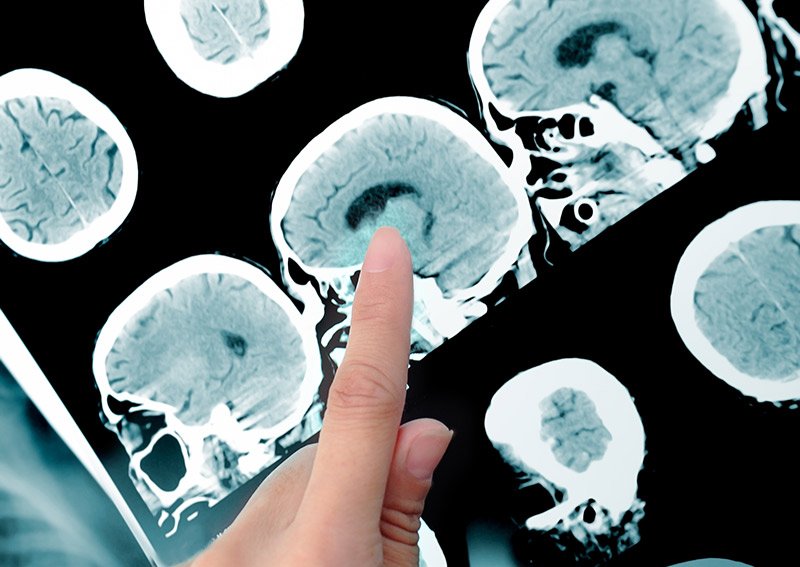
The Amyloid scan uses the tracer Amyvid to detect the brain’s level of amyloid plaque, an indicator of Alzheimer’s disease. A negative scan indicates that the symptoms of cognitive decline are caused by something other than Alzheimer’s, and is useful at ruling out the disease. A positive scan does not necessarily confirm an Alzheimer’s diagnosis, but it can be quite revealing and helpful in managing treatment.
What Can You Detect With A Parkinsons Mri
Generally, you can receive a Parkinson’s diagnosis in a clinical setting, but an MRI can help to assess various aspects of the disease and its progress. In particular, a Parkinson’s MRI can do the following for patients who have or are suspected to have Parkinson’s disease:
- Evaluate tissue loss and how the brain is atrophying
- Check for changes to the basal ganglia region of the brain
- Find out if there are abnormal iron deposits in the basal ganglia or brainstem
- Look at changes to white matter
- Examine the diffusion of restricted tissues in acute infarction and neurodegenerative diseases
- Help to diagnose atypical parkinsonism
- Exclude treatable causes of parkinsonism such as normal pressure hydrocephalus
How Soon Will I Have My Pet Scan Results
PET scans are usually more extensive and detailed than similar tests that are available. Despite this, test results can usually be given within one to two days after the scan.
National Parkinson Foundation: “Should I get a DaT scan or PET scan to confirm my diagnosis of Parkinson’s disease?”
Radiology-Info.org: “PET Scan and Parkinson Disease.”
University of Chicago Medical Center: “How Is Parkinson’s Disease Diagnosed?”
Neurological Imaging In Lakeland Florida
Diagnosing brain and movement disorders is a complex process, mostly dependent on the subjective evaluation of patient symptoms. However, these symptoms often overlap with other neurological conditions, some of which can be treated and/or reversed.
To assist Central Florida physicians in arriving at an accurate diagnosis, Radiology and Imaging Specialists offers the following advanced brain imaging techniques:
New Diagnostic Standards For Parkinsons
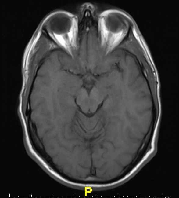
Until recently, the gold-standard checklist for diagnosis came from the U.K.’s Parkinson’s Disease Society Brain Bank. It was a checklist that doctors followed to determine if the symptoms they saw fit the disease. But that’s now considered outdated. Recently, new criteria from the International Parkinson and Movement Disorder Society have come into use. This list reflects the most current understanding of the condition. It allows doctors to reach a more accurate diagnosis so patients can begin treatment at earlier stages.
What Tests Diagnose Parkinson’s Disease
There currently are no tests that can definitively diagnose Parkinson’s Disease. A diagnosis is based on the clinical findings of your physician in combination with your report on the symptoms you are experiencing.??
In situations where an older person presents with the typical features of Parkinson’s and they are responsive to dopamine replacement therapy, there is unlikely to be any benefit to further investigation or imaging.
Computed Tomography Scan Ct Or Cat Scan
A diagnostic imaging procedure that uses a combination of x-rays and computer technology to produce cross-sectional images , both horizontally and vertically, of the body. A CT scan shows detailed images of any part of the body, including the bones, muscles, fat, and organs. CT scans are more detailed than general x-rays.
Physical And Neurological Examination
Your doctor will conduct a physical and neurological examination. This can involve observing your behavior, movements, and mental state and conducting tests or asking you to perform certain exercises.
These are some of the symptoms of Parkinson’s your doctor can determine visually:
- Fewer spontaneous movements or hand gestures
- Reduced frequency of blinking
- Tremors in your hands while they are at rest, often only in one hand
- Hunched posture or forward lean while walking
- Stiff movements
These are some of the exercises your doctor may ask you to do to evaluate your movements, balance, and coordination:
- Opening and closing your fist
- Tapping your fingers, toes, and heels
- Holding your arms out in front of you
- Moving your finger from one point to another
- Rotating your wrists or ankles
- Standing from a chair
Deep Brain Stimulation Benefits for Parkinson’s Can Last at Least 15 Years
What Are The Limitations Of The Test
Currently, DaTscan that is in clinical use is not quantitative, which means that the test is not designed to determine how impaired the dopamine system is – just whether it is or not. This means that the test is not used to tell you whether the disease has progressed over time and is not used to follow a patient’s disease. It also is not used currently as a clinical test to screen for the disease before motor symptoms are evident. Because of these limitations, the search continues for additional measurable indicators, known as biomarkers, to help diagnosis and manage PD.
Tips and Takeaways
- DaTscan is a test that can help in the diagnosis of PD, although in most situations a clinical exam done by a neurologist offers the same information.
- Neurologists are skilled to diagnose PD through a clinical exam. While the exam to some may seem very basic and thus a PD diagnosis subjective or questionable, neurologists are well-trained to assess and diagnose with confidence.
- DaTscan may be useful in distinguishing PD from certain conditions, but not from others, so talk with your neurologist about whether DaTscan would be useful in your specific situation.
- DaTscan is not a test used for monitoring PD progression. It can be used to help clarify a PD diagnosis, but it is not a test you would undergo multiple times during the course of your disease.
Do you have a question or issue that you would like Dr. Gilbert to explore?
Dr. Rebecca Gilbert
How Is Parkinson’s Disease Diagnosed
Your doctor will ask questions about your symptoms and your past health and will do a neurological exam. This exam includes questions and tests that show how well your nerves are working. For example, your doctor will watch how you move. He or she will check your muscle strength and reflexes and will check your vision.
Your doctor also may check your sense of smell and ask you questions about your mood.
In some cases, your doctor will have you try a medicine for Parkinson’s disease. If that medicine helps your symptoms, it may help the doctor find out if you have the disease.
Tests
There are no lab or blood tests that can help your doctor know whether you have Parkinson’s. But you may have tests to help your doctor rule out other diseases that could be causing your symptoms. For example:
- An MRI or CT scan is used to look for signs of a stroke or brain tumor.
- Blood tests check for abnormal thyroid hormone levels or liver damage.
Another type of imaging test, called PET, sometimes may detect low levels of dopamine in the brain. These low levels are a key feature of Parkinson’s. But PET scanning isn’t commonly used to evaluate Parkinson’s. That’s because it’s very expensive, not available in many hospitals, and only used experimentally.
Why Would My Doctor Order A Datscan
Your doctor may order a DaTSCAN™ to confirm or rule out a diagnosis, or to see how a medication is affecting your brain or internal organs. SPECT scans may also be ordered to help diagnose or monitor conditions affecting the bones and the internal organs, especially the heart and the brain, and even to manage and stage cancer.
DaTSCAN™ allows physicians to see which areas of the brain are responding to various stimuli, making it particularly helpful in confirming diagnoses as movement disorders such as Parkinson’s Disease, Parkinsonian Syndrome, and Essential Tremor. DaTSCAN™ can also be helpful in diagnosing and treating traumatic brain injury , attention deficit disorder , attention deficit hyperactivity disorder , and more.
Brain Imaging In Parkinsons Disease
Traditional brain imaging with CT and MRI scans do not show changes in the brain when someone has Parkinson’s disease and are generally not helpful in diagnosis. A new kind of brain scan, called a DaT scan, does show changes in persons with Parkinson’s disease and may someday become an important tool in diagnosing Parkinson’s.
The dopamine transporter, or DaT, scan uses a chemical that labels the dopamine transporter in the area of the brain known as the striatum. Dopamine is a neurochemical that is decreased in persons with Parkinson’s disease.
The dopamine transporter, which moves dopamine in and out of cells, is also decreased in the striatum in persons with Parkinson’s disease and related disorders. The chemical that labels the transporter is injected into the vein and can be imaged by using something called single photon emission computerized tomography, or SPECT scanning. This technique has been registered in the European Union since 2000 for differentiating a diagnosis of essential tremor and a parkinsonian syndrome. It was approved by the Food and Drug Administration in 2011 for this same indication and recently became available at the OHSU Brain Institute.
