Vision Problems Are Common In Parkinsons
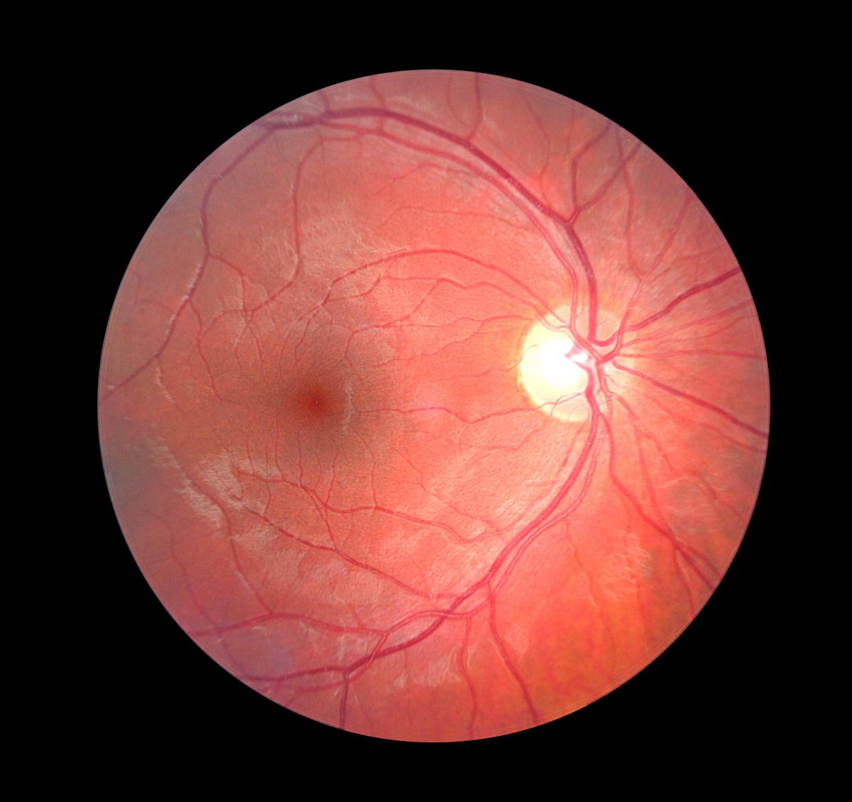
Research has shown that visual symptoms are extraordinarily common in people living with Parkinsons. Visual symptoms may occur due to changes in the front of the eye due to dry eye, changes in the retina , or changes in how our eyes move together. At the same time, many other things can affect vision, including diseases such as age-related macular degeneration, glaucoma, and cataracts, which increase with age. Distinguishing between visual symptoms caused directly by Parkinsons versus one of these other conditions can be difficult.
Visual symptoms related to Parkinsons can be specific: eyes can feel dry, gritty/sandy, and may burn or have redness. You may experience crusting on the lashes, lids that stick together in the morning, sensitivity to light, or dry eye. On the other hand, symptoms can be non- specific: you may notice your vision just isnt what it used to be, and you have difficulty seeing on a rainy night, in dim lighting, or while reading, etc.
Parkinsons Drug Eyed As Treatment For Severe Macular Degeneration
A drug long used to treat Parkinsons disease may benefit patients with a severe form of age-related macular degeneration , a small clinical trial suggests.
One of the leading causes of vision loss in older people is a condition called dry macular degeneration. More than 15% of Americans over age 70 have AMD, and 10% to 15% of those cases go on to develop the more severe wet macular degeneration, which can cause swift and complete vision loss.
Typically, wet AMD is treated with injections of medication into the eye. Most people need several per year to keep the disease from progressing.
But this small, early-stage clinical trial suggests an alternative may be on the horizon: the leading drug used to treat Parkinsons disease, called levodopa.
The trial was an outgrowth of a 2016 study that found Parkinsons patients who took levodopa were less likely to develop macular degeneration.
The study found a relationship between taking levodopa and macular regeneration, said Dr. Robert Snyder, a professor of ophthalmology at the University of Arizona, in Tucson. It delayed the onset of both dry and wet macular degeneration, and reduced the odds of getting wet macular degeneration.
Macular degeneration affects the macula, part of the eye that allows you to see fine detail. Wet AMD happens when abnormal blood vessels grow under the macula often, these blood vessels leak blood and fluid, causing rapid damage.
More information
You May Like: How Long Do People Live With Parkinsons Disease
Macular Degeneration And Vision Loss
Study authors say more than 15 percent of Americans over 70 suffer from macular degeneration. One of the more severe varieties is Neovascular AMD . This type of abnormal blood vessel growth is the result of vascular endothelial growth factor , the buildup of both fluid and blood in the space beneath the retinal.
Less than 20% of AMD patients have this form of the disease, but it accounts for 90 percent of vision loss cases. The normal treatment involves frequent injections to block VEGF, but theyre both expensive and painful for the patient.
Read Also: Can Parkinson Disease Cause Dizziness
Common Parkinsons Disease Drug May Provide Low
PHLADELPHIA Losing your vision can be a terrifying experience. One of the common causes of blindness in seniors is macular degeneration , where leaky blood vessels form under the retina. Treatments for this condition can be both painful and expensive, but a new study says there may be an unlikely solution. A drug for Parkinsons disease is showing promise in reducing the effects of AMD without creating harmful side-effects.
The report finds the drug levodopa is a safe and commonly available medication which reduces a patients need for regular AMD injections. The study continues earlier research which finds patients being treated for various movement disorders, like Parkinsons, are significantly less likely to develop macular degeneration while taking levodopa.
Levodopa has a receptor selectively expressed on pigmented cells. This receptor can be supportive of retinal health and survival, which led to the development of our hypothesis that it may prevent or treat AMD, Robert W. Snyder from the University of Arizona says in a media release.
Want More Practical Articles Like This

You can find much more inourEvery Victory Counts®manual.Its packed with up-to-date information about everything Parkinsons, plus an expanded worksheets and resources section to help you put what youve learned into action.Request your free copy of theEvery Victory Countsmanual by clicking the button below.
Thank you to our 2021 Peak Partners, Adamas, Amneal, Kyowa Kirin, and Sunovion, as well as our Every Victory Counts Gold Sponsor AbbVie Grants, Silver Sponsor Lundbeck, and Bronze Sponsors Supernus and Theravance for helping us provide the Every Victory Counts manual to our community for free.
Also Check: Nyu Langone Parkinson’s Center
Increased Risk For Certain Groups
For individuals over 60 with AMD who also had other medical conditions, the risk of developing Parkinsons disease rose significantly. These medical conditions included:
People with AMD who were also on certain medications were also found to be at a much higher risk for developing Parkinsons disease. These medications include calcium channel blockers and statins .
Thinning Of The Retinal Nerve Fiber Layer
The RNFL is a layer of the retina that is composed of cells called retinal ganglion cells. These cells go on to form the optic nerve, which connects the eye to the brain.
Optical coherence tomography is a test that detects the thickness of the retinal nerve fiber layer. Many OCT clinical studies have found that advanced AMD is associated with a thinner retinal nerve fiber layer.
Doctors have found that measuring RNFL thickness using OCT may be a beneficial tool in following the progression of Parkinsons disease as well as advanced AMD.
Read Also: Big Therapy For Parkinson Disease
Eyes Pupils May Be Window Into Assessing Disease Stage
Prior studies have suggested that Parkinsons patients have a higher tendency for AMD. However, these failed to account for potential additional diseases, or comorbidities.
Now, researchers at the China Medical University Hospital conducted a populationbased retrospective cohort study to assess whether the risk of AMD is elevated in those with Parkinsons, taking into account comorbidities.
They reviewed data from Taiwans National Health Insurance Research Database , specifically the Longitudinal Health Insurance Database 2000 .
In total, they analyzed data from 20,848 individuals, of which 10,424 had AMD and 10,424 did not have AMD . Patients in each group were followed for a mean of 5.66 and 5.48 years, respectively. AMD and non-AMD groups were established from January 1, 2000, to December 31, 2012, to determine the diagnosis of Parkinsons.
The prevalence of comorbidities was significantly higher in the AMD group than in the non-AMD . Medication use, including statins and calcium channel blockers differed slightly between groups.
After adjusting for potential confounders, the data revealed there was a higher risk of developing Parkinsons, both for men and women, in the AMD group than in the non-AMD group.
Specifically, this risk was significantly higher in patients older than 60 and with more than one comorbidity.
AMD is associated with a higher risk of PD with adjustment for sufficient clinical comorbidities and long follow-up time, the researchers concluded.
When To Consult A Doctor
Without treatment, wet macular degeneration is to cause serious vision loss. For this reason, anyone experiencing symptoms of wet macular degeneration should seek advice from a doctor.
Earlier treatments enable doctors to slow the progression of the disease as much as possible.
For some people, the thought of having intravitreal injections may cause concerns. However, a person can ask medical professionals questions about these injections to help alleviate these concerns.
Doctors will also inform people about the potential complications of the procedure. These include:
- discomfort and pain as the anesthetic wears off
- subconjunctival hemorrhage, which refers to eye bleeding
- in rare cases, other complications, such as:
- traumatic cataract
2020 paper explains that intravitreal injections can occur in the operating room or a doctors office.
After administering the anesthesia and cleansing the eye, the medical team will ask the person to look in the direction opposite to where they intend to insert the syringe. After a brief warning, the doctor or surgeon will insert the syringe into the persons eye before injecting the VEGF inhibitors therein. The injections will only take a few moments.
The study also notes that the medical team may irrigate and lubricate the eye after the injection.
Recommended Reading: Slowing Parkinsons Disease Progression
You May Like: Is Parkinson’s Caused By Inflammation
Retinal Pathology In Parkinsons Disease: Implications For Vision And Biomarkers
Study Rationale: Vision problems are not what most of us think of when we think of Parkinsons disease , but there are some physical changes in the retina of people with PD, including an accumulation of the protein alpha-synuclein like that found in the PD brain. It has been suggested that this and/or other eye abnormalities might form the basis of clinical tests that could be used to help diagnose PD or assess whether some PD therapies are working.
Hypothesis: The primary hypothesis is that changes in the retina, including accumulation of alpha-synuclein and/or loss of dopaminergic neurons, will accurately identify people living with PD and that these changes will ultimately allow an ophthalmological diagnosis and assessment of PD severity.
Study Design:We were recently the first to report accumulation of alpha-synuclein in retinal nerve cells of people with PD. This finding needs to be tested in additional people with and without PD to be sure it is a reliable finding. We will also assess the dopaminergic nerve cells of the retina to determine if they are radically depleted, like the dopaminergic neurons in the PD brain. Furthermore, we will assess the overall degree of retinal damage by measuring retinal thickness and numbers of other types of retinal nerve cells, as well as the amount of glial scar tissue that is present.
Advanced Technology For Ocular Degeneration Care
Technology is key in advancing our understanding of neurodegenerative eye diseases at a tissue and cellular level. For example, by using noninvasive, high-resolution optical coherence tomography , we can evaluate the structural changes of the retina in mice and assess how the retina changes along the continuum of the disease and treatment. Early research indicates that OCT measurements can serve as biomarkers for the early recognition and progression of neurological conditions, though further research is needed before such techniques can be fully utilized in a clinical setting.
The UT Southwestern ophthalmology research labs are well-equipped with the cutting-edge instrumentation necessary for elucidating the cause of disease and identifying potential treatments. Our departments instrumentation includes multi-laser flow cytometers with cell-sorting technology for enrichment/analysis studies, focal and full field electroretinography for noninvasive inner/outer retina health measurements, optokinetic reflex monitoring for visual acuity determination, and mass spectrometers for analysis of complex lipid and protein samples.
To discuss your condition with one of our ophthalmology providers, please call or request an appointment.
Recommended Reading: What Is The Difference Between Parkinson’s And Lewy Body Dementia
Microvascular And Choroidal Structure In The Retina
In addition to retina nervous systems, evidence has implicated retinal small vessel plays an important role in structural and functional changes in the retina. Based on the potential risk the role of cerebral small vessel disease for the development of PD plays , some studies demonstrated retinal microvascular changes have been studied retinal capillary plexus vessel density and perfusion density as well as structural changes in PD . Thus, structural changes in retinal microvascular are seen as non-invasive biomarkers for the disease detection.
Considering the common embryologic and anatomic characteristics of retinal vascular with the cerebral circulation, microvascular changes in the retina may correlate with vascular changes in the CNS. The retinal vasculature is a window in vivo non-invasive assessment of microvasculature in the body . In embryology, the ophthalmic artery originates from the internal carotid artery gives off the central retinal artery, providing nutrients and oxygen to the inner layer of the retina. Metabolic waste and carbon dioxide from the retina are excreted into the sinus via the central retinal vein through the superior ocular vein. Central retinal arteries and veins form a terminal branch retinal circulation network on the surface of the retina. In addition, microvasculature of the retina shares similar neurobiology and electrophysiological function with those in CNS .
New Hope For An Amd Treatment

L-DOPA is available worldwide and goes by the generic name of levodopa. Brand names include Sinemet, Parcopa, Atamet, Stalevo, Madopar, and Prolopa.
Despite persistent, dedicated work by scientists, it has been longer than a decade since a new type of drug was developed for AMD, and scientists still do not understand precisely what causes it. AMD remains a chief cause of legal blindness in adults over age 60. While leaving peripheral sight intact , it destroys the center. The resulting black hole makes people unable to drive, read, recognize faces and perform daily tasks.
The only measures currently available to stall and sometimes prevent AMD are the AREDS-proven vitamins for early AMD, and scheduled eye injections for the neovascular form, known as wet AMD.
You May Like: Can Botox Cause Parkinson’s
There Is A Wide Array Of Vision Problems People With Parkinsons May Experience
Here are several common, and a few not-so-common, visual symptoms you may experience:
Blurry vision and difficulty with color vision. Blurry vision may be related to dopamine depletion in the back of the eye and within the visual connections through the brain. This may be partially corrected with dopaminergic medications, though medication effects are usually subtle regarding vision, so you may not notice them.
Visual processing difficulty. This refers to the orientation of lines and edges, as well as depth perception. This can take different forms, including:
- Troubles with peripheral vision: distracted by objects and targets in your peripheral vision
- Difficulties perceiving overlapping objects
- Difficulty copying and recalling figures
- Difficulties detecting whether motion is occurring and in which direction
- Difficulties recognizing faces, facial expressions, and emotions
Dry Eye. Dry eyes are a consequence of decreased blinking and poor production of tears. Dry eye can be worsened by certain medications prescribed for Parkinsons. Dry eye improves with liberal use of artificial tears and good eye/eyelid hygiene. Of note, dry eye doesnt always feel dry! Sometimes it feels like watering, and other times it just feels like blurring or being out of focus.
Results From The Study
The study was based in Taiwan and analyzed more than 20,000 individuals. About half of the people in the study were diagnosed with age-related macular degeneration. The other half did not have AMD. These individuals were followed for about five years.
After adjusting for other factors, researchers found that men and women who had been diagnosed with AMD had a higher risk of developing Parkinsons disease over the 12 years this group was followed.
Don’t Miss: What Is The Pathophysiology Of Parkinson’s Disease
Like Parkinsons Vision Is Linked To The Brain
Vision plays such a critical function that a substantial portion of our brain is made up of pathways that connect our eyes to the visual areas of our brain and the areas that help process this visual information . The primary purpose of the front part of our eyes is to produce the clearest possible image, which is then transmitted to the back part of the eye, called the retina. The retina is made up of nerve cells that communicate via visual pathways using the neurotransmitter dopamine. In addition, we have two eyes with overlapping visual fields, which enables our brain to see the world in three dimensions and process complex visual information.
Guiding Students Toward The Future
Figueroa began working in the McKay Lab as an undergraduate in the summer of 2016 and continues to work there as a research technician, after graduating with a bachelors degree in neuroscience and cognitive science and a minor in physiology and anthropology. After completing two research papers, she plans to turn her attention to medical school applications.
All my students have different paths. My path is to help them get to where theyre going.Brian S. McKay, PhD
Dr. McKay trains high-school to graduate students in laboratory research methods and the concepts behind experimental design as they help him research treatment and prevention of AMD and glaucoma.
My goal is to figure out where the students want to go and help them get there, says Dr. McKay. All my students have different paths. My path is to help them get to where theyre going.
Dr. McKays students follow these paths to a wide range of destinations.
Different students have different goals. In the end, its their life and their decisions, Dr. McKay says. One just graduated from optometry school, one decided shes going to do psychiatry, one is a practicing ophthalmologist in Phoenix, for example.
Don’t Miss: Is Joint Pain A Symptom Of Parkinson’s
Parkinson’s Drug May Help Macular Degeneration
But more research needed to confirm beneficial effects on vision disorder
HealthDay Reporter
THURSDAY, Nov. 12, 2015 — A common Parkinson’s disease medication might hold potential for preventing or treating macular degeneration, the leading cause of vision loss in the elderly, new research suggests.
At this stage, no one is recommending that patients take the drug, levodopa , to thwart eye disease. But the findings are intriguing, researchers said.
“Patients taking L-dopa for any reason are much less likely to develop age-related macular degeneration. If they do, they develop the disease much later in life than those not taking L-dopa,” said study lead author Brian McKay, an associate professor of ophthalmology and vision science at the University of Arizona.
However, the study doesn’t actually prove that levodopa causes a lower incidence of age-related macular degeneration. It only uncovered an association between the two.
Age-related macular degeneration affects about 30 percent of those older than 75, McKay said. It is caused by deterioration of the macula, the center part of the retina, and by affecting vision, it can severely limit the ability to perform everyday activities. Treatments can slow its progression but there is no cure, and it can lead to blindness.
The researchers found that diagnosis of age-related macular degeneration occurred, in general, around age 71. But among those who took levodopa, it occurred much later, at around age 79.
Show Sources
