Neurochemical Deficits In Pdd
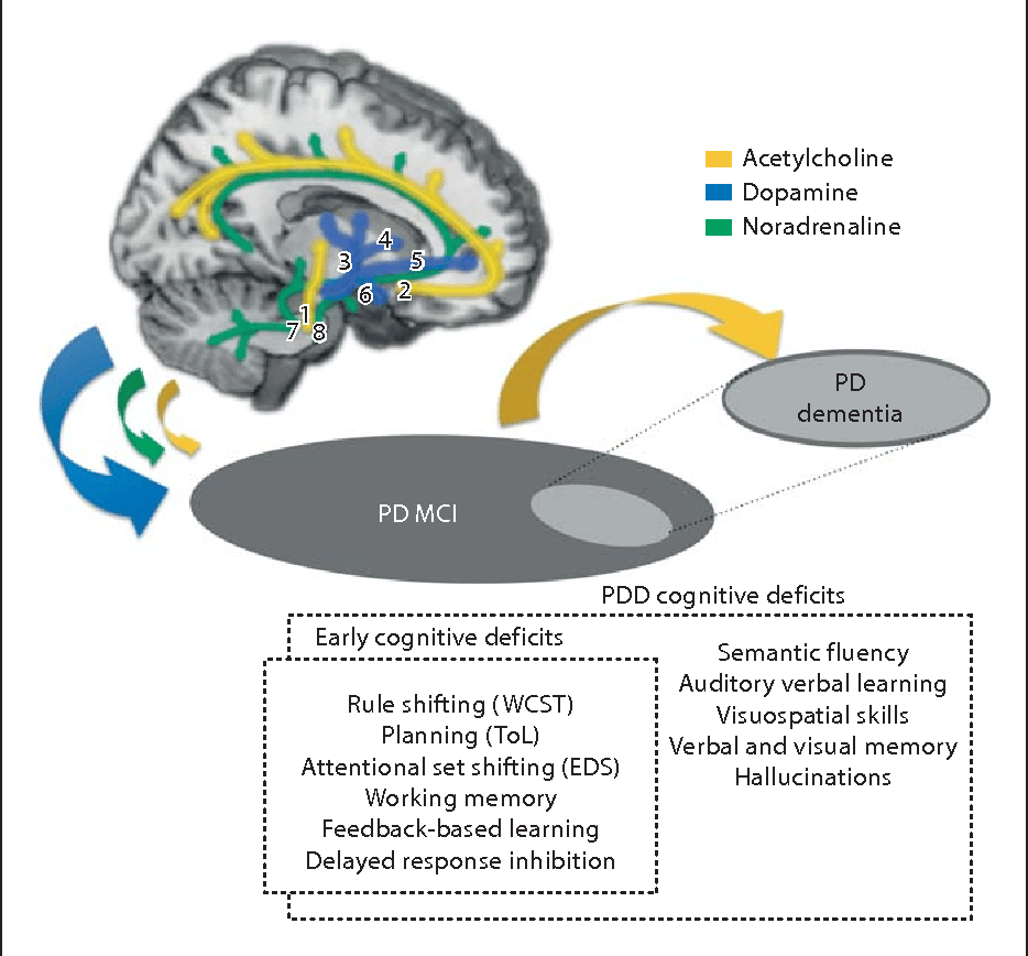
Degeneration of subcortical nuclei in PD leads to dopaminergic, cholinergic, noradrenergic, and serotoninergic deficits. Of them, cholinergic deficits due to degeneration of the nucleus basalis of Meynert have been the most involved in PDD. In early neuropathological studies, PDD patients showed more NMB cholinergic neuronal depletion when compared with AD and non-demented PD.33,34 A greater reduction of choline acetyltransferase activity in frontal and temporal cortex was found in PDD than in PD without dementia.35 Mattila et al reported reduced choline acetyltransferase activity in the hippocampus, prefrontal cortex, and temporal cortex in PD. Reduction in the frontal cortex correlated signicantly with the degree of cognitive impairment.36 Not only pathological studies but also neuroimaging studies have pointed out a role for a cholinergic deficit in cognition in PD. Both PD and PDD have cholinergic neuron decits with vesicular acetylcholine transporter and acetylcholinesterase 37,38 imaging being the decreased VAChT more important and extensive in the cerebral cortex of PDD subjects.39
There are not consistent findings supporting an association between dementia and other monoaminergic systems.
Global Topological Organization Of Functional Brain Networks
The functional brain networks of the three groups exhibited larger > 1 and almost identical 1 compared with those of random networks . Significant group effects were found in the AUCs of Cp, Eloc, Eglob, and Lp . Post hoc testing showed that relative to HC, both PD-M and PD-N had significantly lower Cp , Eloc , and Eglob , and higher Lp there were no significant differences in global metrics between PD-M and PD-N.
Brain regions showing abnormal nodal centrality in functional brain networks in the three groups. In , regions with significant group differences are visualized using the BrainNet viewer . The bar graphs show the post hoc pairwise comparisons with significant differences in nodal degree and nodal efficiency. The y-axes are the area under the curve of the two network parameters and the three groups are color coded as in the key. The x-axis shows the brain regions. Abbreviations: PD, Parkinsons disease PD-M, PD with mild cognitive impairment PD-N, PD with normal cognition HC, healthy control ROL, Rolandic operculum FFG, fusiform gyrus PoCG, postcentral gyrus SMG, supramarginal gyrus PCL, paracentral lobule HES, Heschl gyrus STG, superior temporal gyrus L, left R, right.
How Is Parkinson Disease Treated
Parkinson disease can’t be cured. But there are different therapies that can help control symptoms. Many of the medicines used to treat Parkinson disease help to offset the loss of the chemical dopamine in the brain. Most of these medicines help manage symptoms quite successfully.
A procedure called deep brain stimulation may also be used to treat Parkinson disease. It sends electrical impulses into the brain to help control tremors and twitching movements. Some people may need surgery to manage Parkinson disease symptoms. Surgery may involve destroying small areas of brain tissue responsible for the symptoms. However, these surgeries are rarely done since deep brain stimulation is now available.
Read Also: Is Palsy The Same As Parkinson’s
Network Heat Maps And Individual Optimized Neuromodulation
After transforming our DBS-based cognitive decline circuit into a voxel-wise heat map, we observed that voxels in the ventral anterior STN showed stronger connectivity to our cognitive decline circuit than voxels in the dorsal posterior STN . Using an independent STN DBS validation dataset , we found that intersection of VTAs with our cognitive decline heat map correlated with cognitive decline measured at 1 year post-DBS .
Cognitive decline heat map and validation in an independent cohort. To begin translating our network results into a clinical tool, we generated a heat map where the intensity at each voxels reflects the connectivity of that voxel to our cognitive decline network . In the area of the STN , there is an anterior-posterior and dorsal-ventral gradient. We then tested this heat map in an independent cohort of Parkinsons disease patients with STN DBS and MDRS at baseline and 1 year after DBS . Intersection between VTAs and our cognitive decline heat map was corelated with cognitive decline measured at 1 year .
When patients in our validation cohort were stratified into risk groups based on the degree of VTA overlap with the cognitive decline heat map, there was a significant difference in cognitive decline across groups . Parkinsons disease patients in our high-risk group showed significantly more cognitive decline than patients in our medium risk cohort or low risk cohort .
Clinical Assessments And Neuropsychological Testing

Demographic information we assessed included sex, age at scan, and years of education. Clinical assessments included the Movement Disorders SocietyUnified Parkinson Disease Rating Scale part III motor section both ON and OFF medication, Hoehn and Yahr score , the Geriatric Depression Scale , disease duration, and levodopa equivalent daily dose . Global cognition was assessed with the MoCA . The MoCA is a widely available and quick to administer test with good sensitivity for detecting cognitive impairment in PD . Clinical and cognitive assessments were conducted at time of FBB scan and additional cognitive assessments were performed yearly for 2 years after scan.
Also Check: Parkinson’s Help For Caregivers
Degeneration Of Neurotransmitter Systems
More widespread dopaminergic deficits in the brain
By definition, all patients with PD have a moderate-to-severe loss of dopaminergic neurons in the nigrostriatal projection pathway. More widespread degeneration of dopaminergic terminals in the striatum particularly denervation of dopaminergic terminals in the associative dorsal caudate nucleus occurs in those with PD-MCI than in those with PD without cognitive impairment . However, in patients with PD-MCI, there is relative preservation of other dopaminergic systems in the brain, whilst those with PDD have a considerable loss of the lateral dopaminergic system to frontal, parietal and temporal cortical regions . In healthy individuals, cortical dopamine modulation can boost working memory as well as visuospatial and attentional processing, and promotes cognitive effort,, suggesting a key role for dopamine in cognitive function.
Fig. 2: Neurotransmitter deficits associated with cognitive decline in PD and DLB.
Other pathology
Fbb Image Acquisition And Pre
FBB PET images were acquired at approved PPMI centers in accordance with a standardized FBB imaging protocol . The FBB scans were performed using either a GE or a SIEMENS PET scanner . Images were scanned in a 128×128 matrix size and post reconstruction filter of a Gaussian FWHM 5.0 mm was applied. Participants received the FBB injection as a single intravenous bolus injection consisting of 300 MBq in the antecubital region, followed by a flush of 0.9% sodium chloride to ensure the full radiotracer dose is administered to each participant. Participants rested for 80 min, a 10-min attenuation correction was performed, and then a 4×5-min emission scan was obtained. Participants were at rest and had their heads secured by Velcro during the PET scan.
Table 1 List of the 20 bilateral cortical regions of interest included in the study
We calculated the Calinski-Harabasz index with MATLAB to select the number of clusters we should use as the cut-off solution. Each cluster cut-off solution has a Calinski-Harabasz value which looks to maximize between-cluster variance while minimizing within-cluster variance. The larger the Calinski-Harabasz ratio, the better the solution .
Recommended Reading: How Fast Parkinson’s Progression
How Can Hearing Loss Contribute To Cognitive Decline
It makes sense that hearing trouble can impact cognition. People who do not hear well:
- do not participate in conversation and limit their social and cognitive engagement, which can contribute to cognitive decline.
- use more cognitive energy to decipher the sounds that they are hearing. This leaves less cognitive power for other tasks.
- will have a hard time remembering what they hear.
If those around you tell you that your hearing appears to have diminished, get your hearing evaluated with an audiogram. Improving your hearing with hearing aids could have many positive impacts on your well-being, and potentially improve cognitive functioning as well.
Demographics And Clinical Characteristics
Detailed demographic and clinical information of the 25 PD participants and 30 HCs at time of scan is shown in Table . No data were missing for age, sex, GDS, and years of education for either group and no data were missing for disease duration, MDS-UPDRS-III, and H& Y score for the PD group. Using a two-sample t-test comparing the PD group to HC, the only difference between PD and HC was found in MoCA score, t=2.102 n=55 p=0.043. We did not find any group differences in terms of age, t=0.927 n=55, p=0.36, years of education, t=1.400 n=55 p=0.167, or GDS, t=0.835 n=55 p=0.407. A chi-square test for the nominal variable of sex showed a significant relationship between the two groups in terms of sex ratio, X2 =8.213, p=0.004 however, there is insufficient females in the PD group to further explore this relationship.
Table 2 Participant demographics and clinical characteristics for the Parkinsons disease and healthy control groups at time of Florbetaben scan
Also Check: What Can Cause Tremors Besides Parkinson’s
What Happens In The Brain
The researchers also assessed the impact of ADHD polygenic risk score and amyloid-beta levels on the development of brain abnormalities associated with Alzheimers disease. Specifically, they examined changes in the levels of tau protein and brain morphology during the follow-up period.
The levels of tau protein increase in the brain following the accumulation of beta-amyloid and the progression of Alzheimers disease. As a proxy for tau deposits in the brain, the researchers measured the levels of phosphorylated tau in the cerebrospinal fluid, the colorless fluid that surrounds the brain and the spinal cord.
Individuals with a higher ADHD polygenic risk score who also had higher beta-amyloid levels at baseline showed a greater decline in general cognitive function and memory than either factor alone.
Moreover, a combination of a higher genetic predisposition for ADHD and higher beta-amyloid levels in the brain was associated with the development of brain abnormalities observed in Alzheimers disease.
Specifically, these individuals showed a greater increase in phosphorylated tau levels in the cerebrospinal fluid and degeneration of brain regions involved in cognitive function during the 6-year follow-up period.
Baseline Demographic And Clinical Characteristics
A total of 224/423 patients with PD in the original PPMI cohort were included in this analysis 197 patients were excluded due to having less than 5years of followup or having incomplete year 5 outcome data . In addition, 2 patients were excluded as they were H& Y stage 3 at baseline.
Of the final sample, 58/224 had frequent aggressive dreams at baseline. Baseline demographic and clinical characteristics of patients with and without aggressive dreams are presented in Table 1. The groups did not significantly differ at baseline for demographics, motor impairment, or cognitive function however, patients with aggressive dreams had more severe autonomic symptoms, more frequently reported DEBs, and more often described their dreams as vivid. The symptom profile at followup comparing patients with and without aggressive dreams at baseline is presented in Table 2.
Don’t Miss: New Medicine For Parkinson’s Disease
Predicting Cognitive Decline In Parkinsons Disease Using Fdg
1Department of Human Anatomy and Cell Science, Max Rady College of Medicine, Rady Faculty of Health Sciences, University of Manitoba, Winnipeg, Manitoba, Canada.
2Neuroscience Research Program, Kleysen Institute for Advanced Medicine, Health Sciences Centre, Winnipeg, Manitoba, Canada.
3Department of Neurology, Asan Medical Center, University of Ulsan College of Medicine, Seoul, South Korea.
Address correspondence to: Ji Hyun Ko, Department of Human Anatomy and Cell Science, Max Rady College of Medicine, Rady Faculty of Health Sciences, University of Manitoba, Winnipeg, Manitoba, R3E 0J9, Canada. Email: .
SB and KWP contributed equally to this work.
Find articles byBooth, S.in:JCI |PubMed ||
1Department of Human Anatomy and Cell Science, Max Rady College of Medicine, Rady Faculty of Health Sciences, University of Manitoba, Winnipeg, Manitoba, Canada.
2Neuroscience Research Program, Kleysen Institute for Advanced Medicine, Health Sciences Centre, Winnipeg, Manitoba, Canada.
3Department of Neurology, Asan Medical Center, University of Ulsan College of Medicine, Seoul, South Korea.
Address correspondence to: Ji Hyun Ko, Department of Human Anatomy and Cell Science, Max Rady College of Medicine, Rady Faculty of Health Sciences, University of Manitoba, Winnipeg, Manitoba, R3E 0J9, Canada. Email: .
SB and KWP contributed equally to this work.
Find articles byPark, K.in: |PubMed ||
SB and KWP contributed equally to this work.
Find articles byLee, C.in: |PubMed ||
J Clin Invest.
Hearing Loss & Cognitive Decline

I will start off by discussing one factor that is rarely mentioned in discussions of cognitive decline in PD, but is a highly treatable contributor to cognitive difficulties hearing loss. Abundant research exists that supports the claim that hearing loss impacts cognitive function. The connection was recently highlighted in in the New York Times and in the Wall Street Journal.
One Johns Hopkins research study reviewed thousands of medical claims and demonstrated an association between hearing loss and an increased 10-year risk of dementia, falls, depression and heart attack. Research also suggests that improving hearing with hearing aids can improve cognitive function. One study showed that memory decline slowed in patients who started wearing hearing aids, highlighting the importance of detecting and treating hearing loss early.
Recommended Reading: Are We Close To A Cure For Parkinson’s
Significantly Lower Moca Scores Among Patients With Diabetes Or Prediabetes
No differences were seen between the groups in terms of age, Parkinsons onset and duration, H& Y stage, or in their levodopa equivalent daily dosing , a measure of the total contribution made by each Parkinsons medication.
As expected, blood HbA1c levels were higher in both the diabetes group and prediabetes group compared with those without diabetes .
Notably, MoCA scores were significantly lower indicative of more severe cognitive deficits in both patients with diabetes and prediabetes relative to patients without diabetes .
These differences were maintained even after adjusting for potential influencing factors, including age, sex, disease duration, educational level, H& Y stage, and LEDD. However, there was a considerable overlap in the scores between the three groups, the team noted.
No significant differences in cognitive performance were seen between the diabetes and prediabetes groups. Also, higher HbA1c levels significantly associated with lower MoCA scores in both prediabetic and diabetes-free patients. Patients with diabetes were excluded from this analysis since anti-diabetes medications can affect HbA1c levels.
Further analyses, also adjusting for the same potentially influencing factors, showed that cognitive function declined at a significantly faster rate with aging in Parkinsons patients with diabetes compared with those without this comorbidity.
Path Analysis Of Biomarkers For Cognitive Decline In Early Parkinsons Disease
-
Roles Conceptualization, Data curation, Formal analysis, Investigation, Methodology, Writing review & editing
Affiliation Research and Data Analysis Centre, Brisbane, Queensland, Australia
- Alexandra Gramotnev,
Roles Conceptualization, Data curation, Formal analysis, Investigation, Methodology, Writing review & editing
Affiliations Research and Data Analysis Centre, Brisbane, Queensland, Australia, Sunshine Coast Mind & Neuroscience Thompson Institute, University of the Sunshine Coast, Birtinya, Queensland, Australia
-
Roles Investigation, Writing review & editing
Affiliation School of Health and Behavioural Science, University of the Sunshine Coast, Sippy Downs, Queensland, Australia
Read Also: What Part Of The Body Does Parkinson’s Affect
Connectivity Profiles Of Dbs Effects
Our results align well with a growing literature suggesting that connectivity between DBS sites and brain networks is responsible for DBS induced effects., Further, our results provide further support that a normative connectome can be used to identify these networks. While there is value in obtaining connectivity data from the patients themselves, or using advanced imaging to directly measure remote DBS effects, this is often not possible in clinical settings. In such cases, using a normative connectome averaged across thousands of subjects can serve as useful approximation of the connectivity in each patient. While this approach may sacrifice subject-specific differences in connectivity, this limitation is largely outweighed by the ability to produce robust and reproducible connectivity estimates. Connectomes specific to the population of interest can also be used but are generally of lower quality than normative connectomes and appear to have little impact on results., This normative connectome approach has worked robustly across brain lesions, noninvasive brain stimulation, and DBS. This approach requires no specialized imaging of the patient, only a record of the stimulation location based on routine clinical scans, and thus can be broadly applied in research or clinical settings.
Genetic And Csf Biomarkers
APOE4 genotype and levels of CSF biomarkers were obtained as processed values from the PPMI database. CSF data were available for baseline and follow-up timepoints 1, 2, and 3 of the current study. Briefly, APOE genotyping followed the methods by Livak . Taqman Assays were used per manufacturers protocol to genotype two non-synonymous single nucleotide polymorphisms , rs429358 and rs7412 , in each patient sample in order to distinguish between APOE 2, 3, and 4 alleles. Patients were grouped into those having at least one 4 allele vs. the rest. Genotype was missing for 37 patients .
The measurements of -synuclein, amyloidß42, total -tau, and phosphorylated tau 181 levels in CSF are described in detail in Kang et al. . CSF was collected by standard lumbar puncture. -synuclein, amyloidß42, t-tau, and p-tau were measured by INNO-BIA AlzBio3 immunoassay . -synuclein was measured by enzyme-linked immunosorbent assay. Missing CSF data was 1.7% at baseline, 15.92% at follow-up 1, 17.57% at follow-up 2, and 34.84% at follow-up 3.
You May Like: How Does Parkinson’s Disease Typically Progress
Living With Parkinson Disease
These measures can help you live well with Parkinson disease:
- An exercise routine can help keep muscles flexible and mobile. Exercise also releases natural brain chemicals that can improve emotional well-being.
- High protein meals can benefit your brain chemistry
- Physical, occupational, and speech therapy can help your ability to care for yourself and communicate with others
- If you or your family has questions about Parkinson disease, want information about treatment, or need to find support, you can contact the American Parkinson Disease Association.
