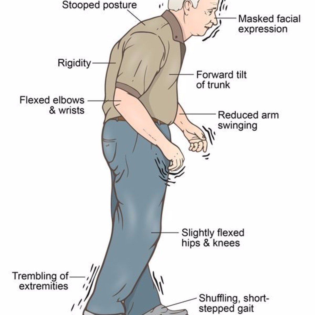
What Causes The Condition
Although there are several recognized risk factors for Parkinsons disease, such as exposure to pesticides, for now, the only confirmed causes of Parkinsons disease are genetic. When Parkinsons disease isnt genetic, experts classify it as idiopathic . That means they dont know exactly why it happens.
Many conditions look like Parkinsons disease but are instead parkinsonism from a specific cause like some psychiatric medications.
Familial Parkinsons disease
Parkinsons disease can have a familial cause, which means you can inherit it from one or both of your parents. However, this only makes up about 10% of all cases.
Experts have linked at least seven different genes to Parkinsons disease. Theyve linked three of those to early-onset of the condition . Some genetic mutations also cause unique, distinguishing features.
Idiopathic Parkinsons disease
Experts believe idiopathic Parkinsons disease happens because of problems with how your body uses a protein called -synuclein . Proteins are chemical molecules that have a very specific shape. When some proteins dont have the correct shape a problem known as protein misfolding your body cant use them and cant break them down.
With nowhere to go, the proteins build up in various places or in certain cells . The buildup of these Lewy bodies causes toxic effects and cell damage.
Induced Parkinsonism
The possible causes are:
Improving Life For Women With Parkinsons Disease
As an audiologist, Sharon Krischer used her skills to help others improve their hearing. But for a long time, she couldnt hear what her own body was telling her.
The mother of three daughters and grandmother of four remembers writing thank you notes one day when her right foot started shaking. It continued happening occasionally, but the inconsistency made Krischer think nothing of it until she broke her opposite leg and the twitch in her right foot returned. This time it wasnt going away.
People living with Parkinsons disease can experience symptoms affecting their movement, as well as other health consequences.
Krischers internist prescribed anti-anxiety medication, but the tremor spread to her right hand. She saw a neurologist who said she had a Parkinsons-like tremor and prescribed an anti-Parkinson drug.
After experiencing hallucinations from the medication, her internist referred her to a movement disorders specialist at University of California, Los Angeles. There, 18 months after first seeing symptoms, she received a diagnosis of Parkinsons disease . She was 57 years old.
The first year is very, very hard if you are a young woman with PD because you dont know how people will react, Krischer said. Its also hard to go from being the caregiver to receiving care, especially if you have children.
You May Like: Levels Of Parkinsons Disease
The Diagnosis Of Idiopathic Parkinsons Disease
The first component to the diagnosis of PD is establishing that the patient has parkinsonism. This is a clinical diagnosis and relies on three key elements: bradykinesia, tremor, and rigidity. Of these, bradykinesia must be present, with at least one of the other two. PD is an asymmetrical condition, so during the clinical assessment, the parkinsonism should be more apparent on one side and may be purely unilateral in early disease . is a classical illustration of parkinsonism, with a description by William Gowers.
A case of Parkinsons disease as described and illustrated by William Gowers: the aspect of the patient is very characteristic. The head is bent forward, and the expression of the face is anxious and fixed, unchanged by any
Read Also: Diagnostic Procedure For Parkinson’s Disease
Benefits Of This Program
- Treat Parkinsons and its symptoms naturally by slightly altering daily habits.
- Every age group can enjoy it and acquire a healthy body in old age.
- Eating healthy food makes the body strong and motivated.
- These foods naturally power up the healing process in the body.
- This program guides how to tackle daily stress and eliminate it during the day.
- Reducing stress gives you proper sleep and improves your mood.
- Taking a healthy diet for a long time will strengthen your bone and muscle.
Imaging The Pharmacology Of Depression In Pd

The prevalence of depression in PD has been reported to range from 10% to 45% . Because Lewy body pathology is known to affect serotonergic and noradrenergic as well as dopaminergic neurotransmission, dysfunction of any or all of these systems would seem to be a reasonable candidate for the functional substrate of depression . To date, functional imaging has failed to demonstrate a correlation between serotonergic dysfunction and depression in PD.
123I–CIT binds with nanomolar affinity to dopamine, noradrenaline, and serotonin transporters. Although striatal uptake of 123I–CIT 24 h after intravenous injection primarily reflects DAT binding, midbrain uptake 1 h after administration reflects serotonin transporter availability . Kim et al. have reported normal brain stem 123I–CIT uptake in PD. They found no difference between uptake in depressed and nondepressed patients and no correlation between radiotracer uptake and Hamilton Depression Rating Scale scores .
Don’t Miss: Is It Parkinson’s Or Something Else
Neurodegeneration With Brain Iron Accumulation
Neurodegeneration with brain iron accumulation patients present with a progressive extrapyramidal syndrome associated with iron deposition in the basal ganglia. The two main syndromes are outlined here, although there are additional syndromes including neuroferritinopathy and aceruloplasminemia. The most common of the NBIA disorders is pantothenate kinase-associated neurodegeneration , resulting from mutations on the PANK2 gene, accounting for 50%. The classic syndrome manifests in early childhood with a combination of pyramidal and extrapyramidal features . PKAN can also rarely present in early adulthood. There are typical MRI findings, with a central hyperintensity with surrounding low signal on T2 images in the globus pallidus, giving the so-called eye-of-the-tiger sign .
The second main type of NBIA is PLA2G6-associated neurodegeneration . When onset occurs in infancy, PLAN causes progressive motor and mental retardation with cerebellar ataxia, seizures, and pyramidal signs. However, onset can occur later in life which leads to an atypical syndrome that may mimic PD, with rest tremor, rigidity, and bradykinesia and a good response to levodopa. However, patients also exhibit additional features including eye-movement abnormalities and pyramidal signs .
Progress Toward Fda Approval
It will be a while yet, however, before Hoque and his researchers can start seeking permission to analyze peoples selfies, or even before neurologists can deploy the five-pronged test that the researchers have developed.
An algorithm will never be 100 percent accurate, Hoque says. What if it makes a mistake? We want to be very careful and follow guidance from the FDA if we want anybody from any part of the world to try this and get an assessment.
Moreover, there is a whole family of movement disorders that are closely related to Parkinsons disease, including ataxia, Huntingtons disease, progressive supranuclear palsy, and multiple dystrophy.
They all share similar symptoms of tremor, but the tremors are very different in nature, Hoque says. However, even expert neurologists find it very, very difficult to distinguish among them.
The researchers have made great progress in detecting Parkinsons disease by automatically analyzing expressions, voice and motor movements. Yet further work is needed to develop algorithms to differentiate how these involuntary tremors differ across other movement disorders, including Ataxia and Huntingtons.
We cant tell that just yet, Hoque says. But we are in a pursuit of differentiating those tremors using AI to prevent the potential harm of misdiagnosis while maximizing benefit.
Also Check: Parkinson’s Wearing Off Symptoms
Idiopathic Basal Ganglia Calcification
This is a heterogenous disease associated with mineral deposition in the basal ganglia, as well as in other brain structures. There is a strong familial component, with causative mutations identified in SCL20A2 and PDGFRB. Patients commonly have a movement disorder, with parkinsonian features of akinesia and rigidity which show a variable response to levodopa. Other features include cognitive impairment, gait disorder, pyramidal signs, and a psychiatric presentation. Imaging is crucial in diagnosis to identify the areas of calcification, with CT imaging being more useful than MRI .
Living With Parkinson Disease
These measures can help you live well with Parkinson disease:
- An exercise routine can help keep muscles flexible and mobile. Exercise also releases natural brain chemicals that can improve emotional well-being.
- High protein meals can benefit your brain chemistry
- Physical, occupational, and speech therapy can help your ability to care for yourself and communicate with others
- If you or your family has questions about Parkinson disease, want information about treatment, or need to find support, you can contact the American Parkinson Disease Association.
Also Check: Keto For Parkinson’s Disease
Nursing Care Plan For Parkinsons Disease 4
Nursing Diagnosis: Disturbed Thought Process related to psychological causes, parkinsonian medications, chronic illness, and depression, secondary to Parkinsons disease as evidenced by memory impairment, distractibility, inability to perform activities, abnormal lab studies, and insomnia.
Desired Outcomes:
- The patient will be able to express understanding of the factors that may produce depressive reactions.
- The patient will use different techniques that will effectively decrease the amount and frequency of depressive reactions.
- The patient will show compliance to the different therapeutic regimens.
Comparison Of The Sl Ll Sllr Area Sim Sisd And Nsim Among The Three Groups
There were no statistically significant differences in SIm_CSF, and SIsd_CSF, nSIm among the three groups. We found significant decreases in SL, cSL, SLLr, cSLLr and Area in the MSA-P group compared to the PD and CGs, a significant increase in SIsd_LN and cSIsd_LN in the MSA-P group compared to the PD group, and a significant decrease in the SIm_LN and nSIm in the MSA-P group compared to the CG. However, no significant difference was found between the MSA-P and PD groups of the SIm_LN and nSIm. The results are shown in Figure 2 and Table 3.
Figure 2. The box-scatter blot of the three groups with upper lower limit, upper lower quartile, and median line. The x-axis represents the three groups using different colors, and the y-axis represents the measured and calculated indexes. corrected short line, cSL, corrected short and long line ration, cSLLr, mean signal intensity of lentiform nucleus, SIm_LN, standard deviation of signal intensity of lentiform nucleus, SIsd_LN, Short line, SL, the ratio of short and long line, SLLr, mean signal intensity of cerebrospinal fluid, SIm_CSF, standard deviation of signal intensity of cerebrospinal fluid SIsd_CSF, long line, LL, Area, normalized mean signal intensity, nSIm, and corrected standard deviation of signal intensity, cSIsd.
Table 3. Comparison of morphological and signal measurement among different group .
You May Like: Anticholinergic Drugs For Parkinson’s
What Causes Parkinsons Disease
The most prominent signs and symptoms of Parkinsons disease occur when nerve cells in the basal ganglia, an area of the brain that controls movement, become impaired and/or die. Normally, these nerve cells, or neurons, produce an important brain chemical known as dopamine. When the neurons die or become impaired, they produce less dopamine, which causes the movement problems associated with the disease. Scientists still do not know what causes the neurons to die.
People with Parkinsons disease also lose the nerve endings that produce norepinephrine, the main chemical messenger of the sympathetic nervous system, which controls many functions of the body, such as heart rate and blood pressure. The loss of norepinephrine might help explain some of the non-movement features of Parkinsons, such as fatigue, irregular blood pressure, decreased movement of food through the digestive tract, and sudden drop in blood pressure when a person stands up from a sitting or lying position.
Many brain cells of people with Parkinsons disease contain Lewy bodies, unusual clumps of the protein alpha-synuclein. Scientists are trying to better understand the normal and abnormal functions of alpha-synuclein and its relationship to genetic mutations that impact Parkinsons andLewy body dementia.
Who Does It Affect

The risk of developing Parkinsons disease naturally increases with age, and the average age at which it starts is 60 years old. Its slightly more common in men or people designated male at birth than in women or people designated female at birth .
While Parkinsons disease is usually age-related, it can happen in adults as young as 20 .
Also Check: How Can I Help My Dad With Parkinson’s
Uniformity Of The Double Measurement Results And Inter
The consistency of the left and right measured data differences between two radiologists was assessed using the ICC. The agreement level definitions based on ICC values were as follows: ICC < 0.3, slight agreement ICC = 0.30.7, moderate agreement ICC > 0.7, good agreement. The ICC values for patients with MSA-P were in good agreement and the best among the three groups, as shown in Table 2.
Table 2. The intraclass correlation coefficient of two radiologists measurement.
Causes And Diagnoses Of Huntingtons Disease
Huntington’s disease is a genetic disorder passed on from parents to children. If a parent has HD, the child has a 50 percent chance of developing it too. Children who dont inherit the gene will not develop the disease and will not pass it along to their children. One to three percent of people with HD have no family history of the disorder.
Read Also: What Are The Five Stages Of Parkinson’s
Single Photon Emission Tomography
The combined pre- and postsynaptic as well as clinical criteria using SPECT imaging method could improve the diagnosis of Parkinsons disease in early stage . The fusion of presynaptic DAT and postsynaptic D2 receptor binding has shown improved diagnostic value in ruling out patients with non-idiopathic parkinsonian syndromes from PD patients .
Dopamine transporter scan
In this test, a radiolabeled tracer, e.g., 123I-ioflupane, is injected into a patients veins, circulates around the body, and gets into the brain. When DAT and dopaminergic neurons reduce in PD and other pre-synaptic parkinsonism diseases, SPECT imaging should take place several hours after the tracer has been administrated. In PD, there is a smaller signal in striatum section of the brain where the ends of the dopamine neurons are meant to be . Indeed, the expression of this protein may reflect the functional dopaminergic neuronal density in striatum part, and its decrease in PD is presumed to be in proportion with severity of the illness .
Fig. 3
DaT scan SPECT images from four patients. Image showing normal comma configuration on the striata bilaterally, with a score of 0. Mild progressive loss of dopamine transporters depicted on the right score of 1, moderate on the left on image score of 2 and severe on the left on image score of 3
123I-ioflupane SPECT imaging
123I-fluopane-CIT
Fig. 4
123I-MIBG scintigraphy
Fig. 5
99mTc-TRODAT-1
Fig. 6Table 2 Some radiotracers used for Parkinsons disease SPECT
Complications Of Parkinsons Disease
Read Also: How Can I Test Myself For Parkinson’s
Why We Need A Laboratory Test For Parkinsons Disease
It is known that the cell loss of PD begins decades before motor symptoms develop and that often certain non-motor symptoms appear first. Therefore, scientists and clinicians are searching for ways to diagnose PD earlier. Diagnosing the disease earlier may allow people with PD to take measures to improve their health earlier, and may be an essential element to developing a neuroprotective medication, a drug that slows down or reverses the nerve damage of PD. It is possible that such a medication would only work at the earliest stages of the disease.
In addition, there are a number of neurologic syndromes that share features of PD. While neurologists are trained to differentiate between these syndromes, researchers are looking for ways to distinguish between different diagnostic possibilities more accurately.
Theres no consensus for a Parkinsons biomarker, making lab tests difficult.
The major obstacle to earlier and more accurate diagnosis of PD, is the current lack of an agreed-upon, simple biomarker for PD. A biomarker is a measurable characteristic in the body that indicates that disease is present. A biomarker can be a lab test, an imaging test, or a clinical test. Common biomarkers include hemoglobin A1c for diabetes, or ejection fraction for heart failure. You can read more about the development of a biomarker for PD in a prior blog.
Pharmacologic Treatment Of Parkinson’s Disease
The goals of treatment are to alleviate symptoms that interfere with the patient’s activities of daily living and to prevent or limit complications as Parkinson’s disease progresses. An additional but still only theoretic goal is to prevent or slow the progression of the disease. If secondary parkinsonism is suspected, treatment should be directed at the identified underlying cause. Treatment for idiopathic Parkinson’s disease should be initiated as soon as the patient’s symptoms begin to interfere with routine activities .
| Drug | |
|---|---|
| 1 to 2 mg twice daily | 13 |
Recommended Reading: How Dopamine Affects Parkinson’s Disease
How Is Parkinsons Disease Managed
Your doctors will tailor your treatment based on your individual circumstances. You will manage your condition best if you have the support of a team, which may include a general practitioner, neurologist, physiotherapist, occupational therapist, psychologist, specialist nurse and dietitian.
While there is no cure for Parkinsons disease, symptoms can be treated with a combination of the following.
