Performance On The Pegboard And Tapping Tasks
All the values given are means for the right and left hand. During the period of 30 seconds, the patient performed 185 unimanual taps. Bimanually, he was able to produce about the same number of taps compared with the unimanual conditions. The number of pegs inserted unimanually was 13 during the period of 30 seconds. Compared with unimanual performance, the number of pegs inserted decreased slightly during the bimanual condition .
In a more complex task, in which the patient placed pegs with one hand, and did tapping with the other, the number of taps clearly decreased . However, the number of pegs inserted remained at the same level compared with the unimanual condition . The results of the pegboard and finger tapping tasks are summarised in table 1.
Performance of pegboard and finger tapping tasks under unimanual, bimanual, and dual task conditions
Blood Sampling And Assessment Of Systemic Oxidative Stress Biomarkers
All participants underwent blood sampling by venepuncture while the neuropsychological assessments and brain MRI evaluations were performed simultaneously. In this study, the percentage of peripheral leucocyte apoptosis was used to assess the oxidative stress. A detailed description of the assessment of leucocyte apoptosis has been presented in previous studies .
The status of leucocyte apoptosis was assessed with APO 2.7-phycoerythrin to identify early and late apoptosis. Positive expression of APO 2.7-PE appears to be restricted to cells undergoing apoptosis. The presence of early apoptotic cells indicated that the apoptotic process was reversible in the early stage, but the presence of late apoptotic cells demonstrated that the cell membrane integrity was disrupted. Leucocytes and their subtypes were analysed according to the intensity of CD45 expression using flow cytometry. Results are expressed as a percentage of specific fluorescence-positive cells. Apoptotic cells were defined as those positive for APO 2.7. A database coordinator monitored all data collection and entry, both of which were checked for any inconsistencies.
Execution Of Simple And Complex Movements
In the RT tasks, the movement times provide information about the speed of execution of simple aimed movements. In general, the MTs in SRT tasks are faster than in CRT tasks both in healthy controls and PD, but MTs do not differ significantly between precued and uncued CRT conditions. Compared with age matched controls, patients with PD have slower MTs. On average, in the SRT task, the MTs of the present case were similar to those of young age matched controls. In contrast, his MTs in the uncued CRT task were even slower than those of the elderly control subjects. In fact our case showed an abnormal pattern of MTs, in that his MTs for the precued CRT task were considerably faster than his MTs for the uncued CRT condition and similar to his MTs for the SRT task . This pattern was not previously seen in either of the young or old control groups or the patients with PD.
Whereas the RT tasks provided fixed start and end points for the movement execution, the movements in the elbow flexion and hand squeeze tasks were self-terminated. Our case was slower in executing aimed movements with a fixed end point as his MTs in the SRT and uncued CRT tasks were slow, although he performed individual elbow flexion and hand squeeze movements at a normal speed. This contrasts with previous findings in parkinsonian patients, in whom it has been suggested that they may have greater difficulties in executing self-terminating movements.
Don’t Miss: End Stage Parkinson Disease Life Expectancy
Pegboard And Finger Tapping
The pegboard and finger tapping tasks were performed as previously described. The pegboard task involved peg placement using the Purdue pegboard. The patient performed the task with the right and the left hand separately, and with both hands simultaneously. On each occasion, the patient had to place as many pegs as possible in a 30 second period. In the finger tapping task, the patient was required to tap a response button repetitively using the index finger for 30 second. The tapping task was performed with the right hand, the left hand, and bimanually. In addition to performing the tapping and pegboard tasks individually, the patient also performed these tasks concurrently, tapping with one hand while placing pegs with the other.
Skin Cancer And Parkinsons Disease

Melanoma;is a type of skin cancer consistently linked to PD.;People who have had melanoma are at an increased risk for PD and people who have PD are at an increased risk of melanoma. Epidemiological studies have shown an increased risk of non-melanoma skin cancers in PD patients as well. Always be sure to talk to your doctor about any skin concerns.
Tips and Takeaways
- Non-motor symptoms such as sweating dysregulation and seborrheic dermatitis can be symptoms of PD
- Seborrheic dermatitis can usually be treated with lifestyle changes and over-the-counter creams. Sometimes prescription-strength creams are necessary
- Although many treatments have been developed for excessive sweating, they have not been tested specifically in people with PD. Discuss with your doctor to find out if any are a possibility for you.
- There is a link between PD and melanoma which you can read about in a prior blog.
- If any symptom is causing you discomfort or interfering with the quality of your daily life, be sure to discuss it with your doctor as it may be something that can be improved with treatment or modifications.
Do you have a question or issue that you would like Dr. Gilbert to explore? Suggest a Topic
Dr. Rebecca Gilbert
APDA Vice President and Chief Scientific Officer
Don’t Miss: Warning Signs Of Parkinsons
Relevance Of The Lesion Network Mapping For Treatment Response
First, we tested whether connectivity with the lesion location was different between patients whose parkinsonism responded to dopaminergic medication compared to those who did not . Specifically, we computed connectivity between each lesion location and an a priori region of interest in the putamen, the site of action for dopaminergic medications . The group differences were investigated using two-sample t-test. To assess specificity, we repeated this analysis on a voxel-wise basis using two-sample t-test.
Second, we tested whether connectivity with our lesion locations might relate to deep brain stimulation response. No patients with lesion-induced parkinsonism underwent DBS. We therefore used a recently published cohort of 95 patients with idiopathic Parkinsons disease who underwent subthalamic nucleus DBS . These patients were used to identify network connections predictive of DBS response . The connectivity of the lesions with the treatment response map was analysed similarly as with the atrophy patterns described in the previous section.
How Is A Diagnosis Made
Because other conditions and medications mimic the symptoms of PD, getting an accurate diagnosis from a physician is important. No single test can confirm a diagnosis of PD, because the symptoms vary from person to person. A thorough history and physical exam should be enough for a diagnosis to be made. Other conditions that have Parkinsons-like symptoms include Parkinsons plus, essential tremor, progressive supranuclear palsy, multi-system atrophy, dystonia, and normal pressure hydrocephalus.
Also Check: Does Parkinson Disease Affect Your Vision
What Is Parkinson’s Disease
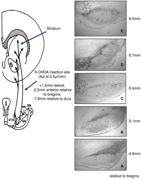
Parkinsons disease is a degenerative, progressive disorder that affects nerve cells in deep parts of the brain called the basal ganglia and the substantia nigra. Nerve cells in the substantia nigra produce the neurotransmitter dopamine and are responsible for relaying messages that plan and control body movement. For reasons not yet understood, the dopamine-producing nerve cells of the substantia nigra begin to die off in some individuals. When 80 percent of dopamine is lost, PD symptoms such as tremor, slowness of movement, stiffness, and balance problems occur.
Body movement is controlled by a complex chain of decisions involving inter-connected groups of nerve cells called ganglia. Information comes to a central area of the brain called the striatum, which works with the substantia nigra to send impulses back and forth from the spinal cord to the brain. The basal ganglia and cerebellum are responsible for ensuring that movement is carried out in a smooth, fluid manner .
The action of dopamine is opposed by another neurotransmitter called acetylcholine. In PD the nerve cells that produce dopamine are dying. The PD symptoms of tremor and stiffness occur when the nerve cells fire and there isn’t enough dopamine to transmit messages. High levels of glutamate, another neurotransmitter, also appear in PD as the body tries to compensate for the lack of dopamine.
Relevance Of The Lesion Network Mapping For Neurodegenerative Parkinsonism
To test whether parkinsonism caused by focal brain lesions shares neuroanatomy with neurodegenerative parkinsonism, we computed functional connectivity between each lesion location causing parkinsonism and maps of brain atrophy from neurodegenerative forms of parkinsonism . For Parkinsons disease, we used a publicly available voxel-wise map of brain atrophy . For PSP and MSA-P, such maps were not available so we used atrophy coordinates from previously published voxel-based morphometry studies . Functional connectivity values between each lesion location and each atrophy map were converted to a normal distribution using Fishers r to z transform. Specificity was assessed in two ways. First, we tested whether lesions causing parkinsonism were more connected to atrophy maps associated with parkinsonism than lesions causing other symptoms. Second, we tested whether lesions causing parkinsonism were more connected to atrophy maps associated with parkinsonism than atrophy maps not associated with parkinsonism . All group comparisons were investigated using two-sample t-tests.
Also Check: Is Hand Shaking A Sign Of Parkinson’s
Preventative Approaches: Targeting The Causes Of Parkinsons Disease
Unfortunately, although some therapies for PD produce a period of recovery for about 5 years, there is a sharp decrease in the beneficial effects of treatments thereafter . Indeed, the best approach would be to understand the relevant triggers of the disease in order to target the physiopathological mechanisms causing the death of dopaminergic neurons. Epidemiological studies have shown that less than 10% of PD cases have a strict familial etiology, while most of them are sporadic and appear to be caused by other factors associated with susceptibility genes . Although these factors are not fully understood, there is a consensus that PD is induced by a combination of age, gender, genetic background, and environmental factors. However, neither of these has, alone, been identified as a leading cause of PD . While the cellular and neurochemical mechanisms underlying PD have remained incompletely understood, what data have been collected point to heavily to mitochondrial dysfunction, oxidative stress, inflammation, and excitotoxicity in the pathogenesis of both familial and sporadic cases of PD .
This evidence suggests that preventing mitochondrial dysfunction can be a key therapeutic goal to achieve as stand-alone or adjunctive therapy against PD.
Lesions In The Substantia Nigra
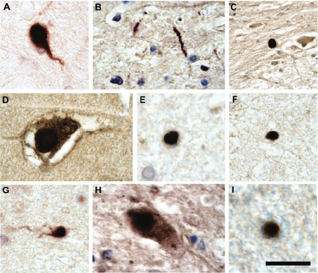
Single, isolated LNs appeared in 4 cases amidst the long, finger-like clusters of heavily melanin-pigmented projection neurons in the posterolateral subnucleus of the pars compacta . No LBs were present and none were seen within the posteromedial, posterosuperior, or magnocellular subnuclei of the pars compacta. The anteromedial, anterointermediate, and anterolateral subnuclei were free of LNs as well as LBs. Three of the 4 individuals were 85 yr of age at death . In the material at our disposal here, no macroscopically discernible depigmentation and no microscopically visible neuromelanin depletion or neuronal loss was evident. The melanoneurons of the paranigral nucleus, parabrachial pigmented nucleus, and perirubral formation still were free of lesions.
Recommended Reading: How Does A Person Die From Parkinson’s
Localizing Parkinsonism Based On Focal Brain Lesions
1Athinoula A. Martinos Center for Biomedical Imaging, Massachusetts General Hospital, Charlestown, MA, USA
2Berenson-Allen Center for Noninvasive Brain Stimulation, Beth Israel Deaconess Medical Center, Boston, MA, USA
3Harvard Medical School, Boston, MA, USA
4Department of Neurology, University of Turku, Turku, Finland
5Division of Clinical Neurosciences, Turku University Hospital, Turku, Finland
What Are The Causes

The cause of Parkinson’s is largely unknown. Scientists are currently investigating the role that genetics, environmental factors, and the natural process of aging have on cell death and PD.
There are also secondary forms of PD that are caused by medications such as haloperidol , reserpine , and metoclopramide .
Don’t Miss: What Toxins Can Cause Parkinson’s Disease
Value Of Accurate Diagnosis
An accurate diagnosis in a person with suspected Parkinsons has an important bearing on prognosis especially in those with younger-onset diagnosis. People with Parkinsons will have a longer life expectancy than those with MSA or PSP and will respond better to dopaminergic medication. It should also be differentiated from other conditions presenting with tremor such as a postural and action tremor similar to that seen in essential tremor. Other causes of Parkinsonism and Parkinson’s-Plus syndromes include multiple cerebral infarctions. Differential diagnosis can also be difficult in elderly people since extrapyramidal symptoms and signs are common.
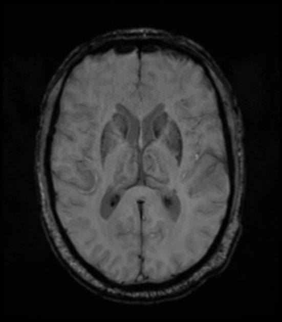
Clinical Characteristics In Pd Patients With White Matter Lesions
The clinical characteristics of patients with idiopathic PD are presented in Table;. A total of 146 patients with PD, including 64 males and 82 females, were enrolled in this study. The mean age of the participants was 63.68years. The percentages of hypertension, diabetes mellitus, hypercholesterolemia and smoking were 40, 13, 21.2 and 2.7%, respectively. The percentage of medications use that possible interfere with cognitive performance, which defined as Benzodiazepines , was 35.6%. The mean total scores of UPDRS were 38.54, the mean stages in modified HYSS, the SE-ADLS, and MMSE were 1.94, 82.96, and 17.91, respectively. The mean grading of the PWMH and DWMH were 1.16 and 1.12, respectively.
Table 1 Clinical characteristics in PD
Read Also: How To Stop Parkinson’s From Progressing
Flex And Squeeze Tasks
Flex and squeeze tasks were performed as previously described. The patient sat with his forearm flexed at 90°, resting on a manipulandum, with his hand holding a vertical grip containing a strain gauge. The patient performed two types of movement, a 15° flexion of the elbow and an isometric 30 Newton hand squeeze. Three conditions were assessed: simple flexion performed alone, the two movements performed simultaneously, and finally the two movements performed sequentially in the order squeeze then flex. For the sequential task, the inter-onset latency between the onsets of the squeeze and flex was also determined.
Performance Of The Flex And Squeeze Tasks
Movement times for the two tasks performed separately are for the right hand. In the simultaneous and sequential tasks, hand squeeze was performed with the left hand, and elbow flexion with the right hand. In the sequential task, hand squeeze was performed first, followed by elbow flexion. The movement time for squeeze alone was 158 ms. Hand squeeze performance slowed down to some extent when performed simultaneously with elbow flexion . The movement time for elbow flexion alone was 209 ms. Elbow flexion performance remained at about the same level when performed simultaneously with hand squeeze . In the sequential task, performance of elbow flexion clearly slowed down . The results of the flex and squeeze tasks are summarised in table 2.
Contingent negative variation.
Also Check: Does Medicare Cover Dbs For Parkinson’s
When Are Lesion Surgeries Indicated In The Treatment Of Parkinson Disease
Lesion surgeries involve the destruction of targeted areas of the brain to control the symptoms of Parkinson disease. Lesion surgeries for Parkinson disease have largely been replaced by deep brain stimulation . During neuroablation, a specific deep brain target is destroyed by thermocoagulation. A radiofrequency generator is used most commonly to heat the lesioning electrode tip to the prescribed temperature in a controlled fashion.
Contributions Of Research Using The Mptp Monkey Model To Understanding The Pathophysiology Of Pd
Using the MPTP monkey model of PD electrophysiological recordings in the STN, globus pallidus internus and globus pallidus externus were performed by several groups . Compared to the naive state these studies reported changes in mean discharge rate with increased rates in the STN and GPi and decreased rates in GPe as well as a loss of specificity and increased number of cells responsive to passive manipulation . Based on these findings a rate model of PD was proposed in which the direct pathway, projections from the putamen to GPi, was underactive and the indirect pathway, projections from putamen to GPe, was overactive . This model hypothesized that mean discharge rates in the STN would therefore be increased in PD, leading to excessive activation of the GPi and suppression of thalamocortical activity leading in turn to the manifestation of Parkinsons motor signs . A seminal test of this hypothesis was published in Science in 1990 .
During this time the number of pallidotomies grew and given PD is a progressive disorder bilateral pallidotomies were required for patients with disease affecting both sides of the body. Complications with bilateral pallidotomy, however, were too frequent, with some reporting cognitive changes, gait disorders, worsening PD, and/or hypophonia . An alternative approach was needed, and deep brain stimulation for PD, developed by Alim Louis Benabid, was brought to the operating room .
Recommended Reading: How To Know If You Have Parkinson’s Disease
Significance Of Brain Lesions In Parkinson Disease Dementia And Lewy Body Dementia
Log in to MyKarger to check if you already have access to this content.
Buy a Karger Article Bundle and profit from a discount!
If you would like to redeem your KAB credit, please log in.
Save over 20%
- Rent for 48h to view
- Buy Cloud Access for unlimited viewing via different devices
- Synchronizing in the ReadCube Cloud
- Printing and saving restrictions apply
USD 8.50
- Immediate access to all parts of this book
- Cover-to-cover formats may be available
- Unlimited re-access via MyKarger
The final prices may differ from the prices shown due to specifics of VAT rules.
Lesions In The Olfactory Bulb Olfactory Tract And Anterior Olfactory Nucleus

In 16 cases, one or more anterior olfactory structures displayed PD-related pathology . Elongated rather than thick-caliber LNs surpassed small LBs numerically. The lesions occurred in layers VVI of the olb, which is characterized by a concentric eight-layered lamination pattern , as well as in the otr and cellular islands of the aon harbored by the otr. Exceptionally dense accumulations of LNs interspersed by LBs could be seen in the aon, which contained 2 types of neurons: both presented with LBs and both contained lipofuscin granules . Where portions of above-mentioned anterior olfactory entities from both hemispheres were intact , the lesions were bilateral in 1 case . In 1 case , the olb and otr were the only sites sustaining damage in addition to the dmo . In no instance were anterior olfactory structures affected to the exclusion of all other nuclear grays examined . The olfactory tubercle, piriform cortex, and periamygdaloid cortex did not contain LNs and/or LBs .
You May Like: Does Parkinson’s Affect Balance
