Mechanisms Of Action In Reducing Levodopa
Pallidal stimulation
Restoration of the thalamocortical activity by suppression of the inhibitory output from the pallidum to the ventrolateral thalamus is the suspected mechanism for motor improvement underpinning GPi DBS, however, the cellular mechanisms of high-frequency stimulation are still unknown. The mechanism of GPi DBS in reducing dyskinesia is also not completely understood. The current views of the BG physiology suggest that inhibition of ventral GPi activity should induce dyskinesia, however, lesioning of the ventral pallidum provides relief of dyskinesia . One of the possible justifications for this apparent paradoxical response is that LID may be more correlated with an abnormal pattern than with the direction and intensity of the neuronal activity within the GPi . Surgical modification of this patterned activity might be accomplished by lesioning or with DBS . Dyskinesia might also arise from an abnormal balance of activity within different functional zones of the nucleus and stimulation may suppress this abnormal activity . Finally, the anti-dyskinetic effect of GPi DBS maybe mediated through effects on the subthalamopallidal tract, which projects to the dorsal GP externus and GPi. Dorsal GPi stimulation might inhibit this projection and would be expected to improve PD symptoms and induce dyskinesia .
STN stimulation
What Happens During Surgery
For stage 1, implanting the electrodes in the brain, the entire process lasts 4 to 6 hours. The surgery generally lasts 3 to 4 hours.
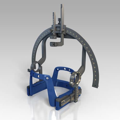
Step 1: attach stereotactic frameThe procedure is performed stereotactically, which requires attaching a frame to your head. While you are seated, the frame is temporarily positioned on your head with Velcro straps. The four pin sites are injected with local anesthesia to minimize discomfort. You will feel some pressure as the pins are tightened .
Step 2: MRI or CT scanYou will then have an imaging scan, using either CT or MRI. A box-shaped localizing device is placed over the top of the frame. Markers in the box show up on the scan and help pinpoint the exact three-dimensional coordinates of the target area within the brain. The surgeon uses the MRI / CT scans and special computer software to plan the trajectory of the electrode.
Step 3: skin and skull incisionYou will be taken to the operating room. You will lie on the table and the stereotactic head frame will be secured. This prevents any small movements of your head while inserting the electrodes. You will remain awake during surgery. Light sedation is given to make you more comfortable during the initial skin incision, but then stopped so that you can talk to the doctors and perform tasks.
What Is Parkinsons Disease Surgery
Parkinsons disease surgery is a brain operation called deep brain stimulation . The surgery is also used to treat epilepsy, obsessive-compulsive disorder and a condition called essential tremor. DBS is widely considered one of the most significant neurological breakthroughs in recent history, posing a potential treatment for major depressive disorder, stroke recovery and addiction. Parkinsons disease brain surgery aims to interrupt problematic electrical signals from targeted areas in the brain and reduce PD symptoms.
You May Like: How To Help A Person With Parkinson Disease
Don’t Miss: What’s The Signs Of Parkinson’s Disease
Figure 81structures Of The Basal Ganglia262
Experimental studies using the MPTP primate model showed increased cellular activity in the STN, and lesions or stimulation of the STN can reverse the cardinal features of parkinsonism., However, surgeons were reluctant to lesion the STN in humans because of the risk of inducing hemiballismus. It was then shown that electrical stimulation of the STN- produced dramatic improvement in parkinsonian symptoms in . STN-DBS has since become the most widely undertaken surgical procedure for PD.
Surgical techniques vary between centres, but it is generally performed in three stages: radiological localisation, physiological localisation, and then either an ablation or a stimulation procedure.
Radiological localisation involves the rigid fixation to the skull under local anaesthesia of a stereotactic base ring onto which a fiducial array can be mounted. In the past, ventriculography was the radiological technique used, but this has been largely replaced by CT and MRI. It is now possible to identify most of the targets on MRI, and their position in stereotactic space is calculated using sophisticated computer programs.
In view of the relative safety of stimulation procedures compared with lesioning, most surgery for people with today uses the former approach. The felt therefore that it should confine its recommendations to STN, GPi and thalamic stimulation.
What Are The Risks

As with any surgery, there are risks associated with deep brain stimulation. If you have DBS, you have about a 3% risk of seizure, infection and confusion. Parkinsons disease deep brain stimulation also carries a small chance of stroke. Once the neurotransmitter device is turned on, you may experience side-effects such as numbness, muscle tightness, tingling sensations balance problems or mood changes these can be managed with help from your doctor.
Your doctor will work with you to assess your Parkinsons disease and surgery eligibility and discuss the advantages and disadvantages of this treatment route. If you have any questions, do not hesitate to run them past your treatment provider or call the National Parkinsons Foundation helpline on 1-800-4PD-INFO for more information.
APA ReferenceSmith, E. . How Effective is Brain Surgery for Parkinsons Disease?, HealthyPlace. Retrieved on 2021, August 26 from https://www.healthyplace.com/parkinsons-disease/treatment/how-effective-is-brain-surgery-for-parkinsons-disease
Read Also: Is There A Cure For Parkinson’s Disease
Evaluation Of Patients For Stereotactic Surgery
Good surgical outcomes from stereotactic surgery for Parkinson’s disease begin with careful patient selection and end with attentive, detail-oriented postoperative care. The authors believe that this level of care is best provided by a multidisciplinary team comprising a movement disorder neurologist, a neurosurgeon who is well versed in stereotactic technique, a neurophysiologist, a psychiatrist, and a neuropsychologist. Additional support from neuroradiology and rehabilitation medicine is also important. But, as a European-based survey revealed, having the proper clinical support is only one of the challenges that stereotactic surgery for PD faces. There are also issues of regulatory, technical, scientific, and intellectual property rules as well as public perception challenges that have to be overcome.
In addition to being medically evaluated, patients are evaluated for surgery in the movement disorder centers by a neurologist, a neurosurgeon/neuro-radiotherapist, and a psychiatrist/psychologist.
A neurologist with expertise in movement disorders evaluates the patient to assure they are a good candidate for a successful subthalamic nucleus deep brain stimulation to confirm a diagnosis of idiopathic PD, positive response to levodopa, absence of atypical parkinsonian features, and assess the advancement of disease as unmanageable with dopaminergic medications. Other requirements include:
Benefits And Side Effects
FUS is a less invasive approach than others doctors do not need to make incisions or holes in the skull, meaning there is a lower risk of infections and bleeds. The procedure is also incredibly precise, limiting damage to surrounding parts of the brain.
Currently, a doctor may only use FUS for the treatment of tremors due to Parkinsons. Clinical trials are investigating the potential benefits of FUS in targeting other areas of the brain to help treat symptoms such as dyskinesias.
A person can usually return home the same day as the procedure, but they may experience some side effects such as numbness in the face or arms, poor balance, and speech and swallowing problems. However, these side effects tend to be temporary.
You May Like: Boxing And Parkinsons Disease
Also Check: Can You Have Parkinson’s Without Tremors
What Benefits Does The Procedure Offer
DBS is not a cure for Parkinsons, but it may help control motor symptoms while allowing a reduction in levodopa dose. This can help reduce dyskinesias and reduce off time. DBS does not usually increase the peak benefits derived from a dose of levodopa the best levodopa response before DBS is a good indicator of the best response after DBS. But it can help extend the amount of on time without dyskinesias, which may significantly increase quality of life.
DBS does not provide most patients benefit for their non-motor symptoms, such as depression, sleep disturbance, or anxiety. DBS also does not usually improve postural instability or walking problems. If a symptom you have does not respond to levodopa, it is not likely to respond to DBS.
Historical Perspective Of Deep Brain Stimulation
Prior to the discovery of levodopa, surgical interventions were the most efficacious treatment for PD symptoms, but primarily focused on the reduction of bothersome tremor. Early approaches targeted the pyramidal tracts, with lesioning either at the point of origin in the cortex or the descending pathways through the brainstem and cervical spinal cord . Although tremor was reliably improved following surgery, hemiparesis was an inevitable consequence. However, in 1952, Dr. Irving Cooper inadvertently interrupted the anterior choroidal artery while performing a mesencephalic pedunculotomy in a patient with PD. Ligation of the vessel was required, though what resulted was a serendipitous reduction in rigidity and tremor with preservation of motor and sensory function. Cooper reasoned the favorable outcomes were due to infarction of the medial globus pallidus. An expansion of ablative stereotactic surgery followed, aided by the earlier development of the stereotactic frame and methods of targeting deep brain structures, including the basal ganglia and thalamus. However, the success of these approaches was limited, partly because of inaccurate, imprecise, and inconsistent targeting. Moreover, intentionally created bilateral brain lesions frequently led to irreversible deficits in speech, swallowing, and cognition.
Also Check: How To Support Someone With Parkinsons Disease
Read Also: What Does Parkinson’s Stiffness Feel Like
What You Need To Know
- Surgeons implant one or more small wires in the brain during a surgical procedure.
- The leads receive mild electrical stimulation from a small pulse generator implanted in the chest.
- Proper patient selection, precise placement of the electrodes and adjustment of the pulse generator are essential for successful DBS surgery.
- DBS does not fully resolve the symptoms of PD or other conditions, but it can decrease a patients need for medications and improve quality of life.
What Are The Results
Successful DBS is related to 1) appropriate patient selection, 2) appropriate selection of the brain area for stimulation, 3) precise positioning of the electrode during surgery, and 4) experienced programming and medication management.
For Parkinson’s disease, DBS of the subthalamic nucleus improves the symptoms of slowness, tremor, and rigidity in about 70% of patients . Most people are able to reduce their medications and lessen their side effects, including dyskinesias. It has also been shown to be superior in long term management of symptoms than medications .
For essential tremor, DBS of the thalamus may significantly reduce hand tremor in 60 to 90% of patients and may improve head and voice tremor.
DBS of the globus pallidus is most useful in treatment of dyskinesias , dystonias, as well as other tremors. For dystonia, DBS of the GPi may be the only effective treatment for debilitating symptoms. Though recent studies show little difference between GPi-DBS and STN-DBS.
Patients report other benefits of DBS. For example, better sleep, more involvement in physical activity, and improved quality of life .
Research suggests that DBS may “protect” or slow the Parkinson’s disease process .
You May Like: Special Walker For Parkinson’s Patients
How Should I Care For The Surgical Area Once I Am Home
- Your stitches or staples will be removed 10 to 14 days after surgery.
- Each of the four pin sites should be kept covered with band aids until they are dry. You will be able to wash your head with a damp cloth, avoiding the surgical area.
- You may only shampoo your hair the day after your stitches or staples are removed, but only very gently.
- You should not scratch or irritate the wound areas.
Practical Issues Selection Of The Surgical Target Technique And Programing

Table 1 shows a summary of points that need to be considered when indicating these DBS techniques.
Numerous studies have demonstrated the effectiveness of STN DBS in controlling the appendicular motor signs of PD, however, this procedure is not considered to have as much of a direct effect on the intensity of LID. The anti-dyskinetic effect of STN DBS have been hypothesized to be related to allowing reduction of dopaminergic drug dosages, with consequent improvement in side effects, including LID. The persistence or worsening of LID after STN DBS is common and is, in fact, indicative of the necessity to reduce the dose of levodopa . Therefore, the si ne qua non-condition for reduction of LID when STN DBS is considered, is its capacity to enable a reduction of levodopa dosage. If, however, an adequate response of motor symptoms does not occur post-operatively, dyskinesia will remain unchanged. Of importance, STN stimulation not uncommonly induces contralateral dyskinesia, which may be persistent, and in some cases lead to the implantation of rescue GPi leads.
In the case of a patient in whom, in addition to motor signs of parkinsonism, medication side effects other than dyskinesia are a primary source of disability , STN DBS may be more desirable.
Post-operative programing: GPi DBS
Post-operative programing: STN DBS
Don’t Miss: How Does A Person Get Parkinson’s Disease
How Deep Brain Stimulation Works
Exactly how DBS works is not completely understood, but many experts believe it regulates abnormal electrical signaling patterns in the brain. To control normal movement and other functions, brain cells communicate with each other using electrical signals. In Parkinsons disease, these signals become irregular and uncoordinated, which leads to motor symptoms. DBS may interrupt the irregular signaling patterns so cells can communicate more smoothly and symptoms lessen.
You May Like: Parkinsonism Is Characterized By The Loss Of
What Is Deep Brain Stimulation
Deep brain stimulation is a surgical procedure that involves implanting electrodes in the brain, which deliver electrical impulses that block or change the abnormal activity that cause symptoms.
The deep brain stimulation system consists of four parts:
- Leads that end in electrodes that are implanted in the brain
- A small pacemaker-like device, called a pulse generator, that creates the electrical pulses
- Extension leads that carry electrical pulses from the device and are attached to the leads implanted in the brain
- Hand-held programmer device that adjusts the devices signals and can turn the device off and on.
In deep brain stimulation, electrodes are placed in the targeted areas of the brain. The electrodes are connected by wires to a type of pacemaker device placed under the skin of the chest below the collarbone.
Once activated, the pulse generator sends continuous electrical pulses to the target areas in the brain, modifying the brain circuits in that area of the brain. The deep brain stimulation system operates much the same way as a pacemaker for the heart. In fact, deep brain stimulation is referred to as the pacemaker for the brain.
Don’t Miss: How Hereditary Is Parkinson’s Disease
Advanced Medicinal Products For Parkinson’s Disease
For the last couple of decades several new and exciting technologies have been investigated to address a plethora of brain diseases, including Parkinson’s disease , with positive experimental results. However, these advances have lead to the development of medical tourism to obtain unproven or disapproved therapies .
Some mostly experimental and unapproved therapies include:
-
grafting of stem cell-derived dopaminergic neurons into the striatum
-
transfection of cells in the striatum with dopamine gene
-
neurorestorative therapies, which use gene- or cell-based therapies to release certain growth factor
Those transplantation techniques are a potential treatment for PD for the following reasons:
-
The neuronal degeneration is site- and type-specific
-
The target area is well defined
-
Postsynaptic receptors are relatively intact
-
The neurons provide tonic stimulation of the receptors and appear to serve a modulatory function
In double-blind studies, neither transplantation of autologous adrenal medullary cells nor transplantation of fetal porcine cells were effective thus, both were abandoned. Although open-label studies of fetal dopaminergic cell transplantation yielded promising results, 3 randomized, double-blind, sham-surgerycontrolled studies found no benefit. In addition, some patients receiving these transplants developed a potentially disabling form of dyskinesia that persisted even after the withdrawal of levodopa.
References
What Happens After Surgery
After surgery, you may take your regular dose of Parkinsons medication immediately. You are kept overnight for monitoring and observation. Most patients are discharged home the next day.
During the recovery time after implanting the electrodes, you may feel better than normal. Brain swelling around the electrode tip causes a lesion effect that lasts a couple days to weeks. This temporary effect is a good predictor of your outcome once the stimulator is implanted and programmed.
About a week later, you will return to the hospital for outpatient surgery to implant the stimulator in the chest/abdomen. This surgery is performed under general anesthesia and takes about an hour. Patients go home the same day.
Step 7: implant the stimulator You will be taken to the OR and put to sleep with general anesthesia. A portion of the scalp incision is reopened to access the leads. A small incision is made near the collarbone and the neurostimulator is implanted under the skin. The lead is attached to an extension wire that is passed under the skin of the scalp, down the neck, to the stimulator/battery in the chest or abdomen. The device will be visible as a small bulge under the skin, but it is usually not seen under clothes.
You should avoid arm movements over your shoulder and excessive stretching of your neck while the incisions heal. Pain at the incision sites can be managed with medication.
Read Also: What Age Parkinsons Disease
You May Like: Does Parkinson’s Affect Eyesight
Life After Dbs Surgery
Once the neurotransmitter has been programmed, you are given a handheld controller to make adjustments.
With the controller, you can turn the simulator on or off, select the signal strength, and move across different program types.
If your DBS neurotransmitter has a rechargeable battery, then it will take about two hours for the device to recharge completely.
Make sure to carry your Implanted Device Identification card if you are traveling by air, as Airport Security will detect the device.
