Lipid Raft Anomalies And Neuropathology
Taking into account the variety of functions that have been related to lipid rafts, it is conceivable that alterations in the lipid composition of these membrane sites may have important consequences for neuronal stability and functionality. Recently, it has been established that a parameter promoting lipid unbalance of lipid rafts is just the consequence of brain ageing progression . Cerebral ageing is a complex process where multiple factors intervene, including mitochondrial impairment, metabolic alterations, a progressive neuronal detriment, loss of neuronal plasticity, increase of oxidative stress, and the accumulation of toxic protein aggregates . For instance, the most relevant aged-related neuropathologies, i.e., Alzheimer disease , Parkinsons disease , dementia with Lewy bodies , Huntington disease, and frontotemporal dementia are generally characterized by abnormal protein oligomerization and aggregate formation that accumulates in different brain areas, contributing to the neuropathology . Fig. 18.1 illustrates a potential scenario of the molecular events occurring in lipid rafts during the progression of neurodegeneration .
Figure 18.2. Schematics of the potential neuropathological lipid changes in lipid rafts.
Table 18.2. Lipid alterations in neuronal lipid rafts associated with Alzheimer disease and synucleopathies .
| Neuronal tissue |
|---|
Anne Bertolotti, in, 2018
Ali Shariati, … Ali Hassanzadeh, in, 2020
Who Gets Als And Ms
MS is estimated to affect over 2.3 million people worldwide, with approximately 1 million of them in the United States.
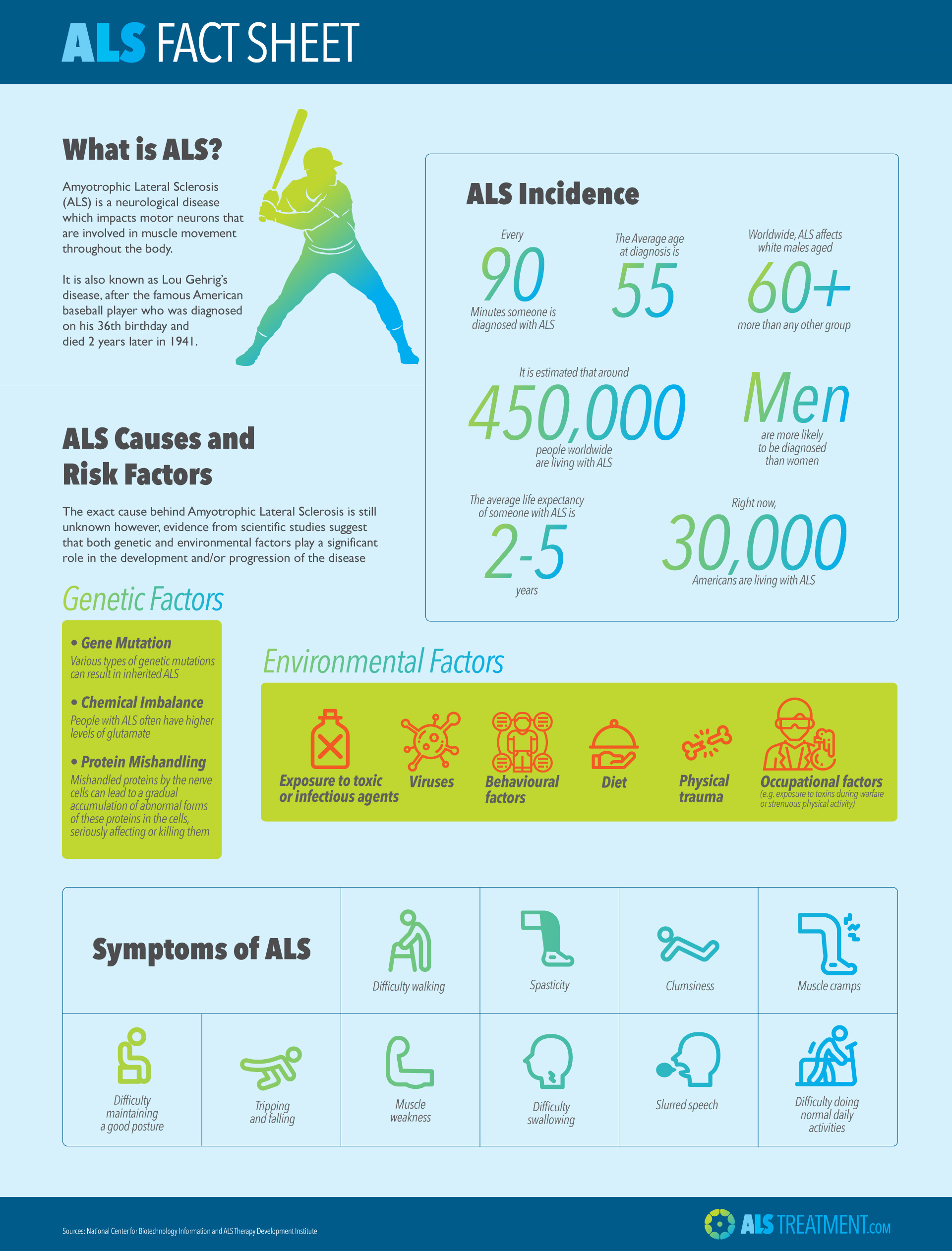
Around 30,000 people in the United States live with ALS, according to the Hospital for Special Surgery. Over 5,600 people in the country are diagnosed with ALS every year.
There are several risk factors that may affect who develops ALS and MS.
Als As A Disease Of Rna Quality Control
Although ALS research was dominated for many years by the apparent connection between SOD1 misfolding and FALS, the recent discovery of two additional genes, mutations which appear to be associated with both familial disease and increased risk for sporadic ALS, has caused a rethinking of the molecular etiology of most forms of the disorder. In contrast to the antioxidant enzymatic activity of SOD1 and its tendency to form pathological aggregates, these two new genes, TDP-43 and FUS/TLS, encode primarily nuclear proteins that function in RNA biology. As we shall see, they not only raise the possibility that ALS is an RNA-based disease, they also suggest that a broader view of the concept of proteostasis may be valuable for developing new approaches to its treatment.
Also Check: What Is The Life Expectancy Of Someone With Parkinson’s Disease
Difference In Parkinsons Disease And Als Diagnosis And Treatment
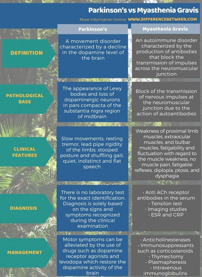
There is currently no specific test that can be performed to directly diagnose Parkinsons disease, but an array of different tests can help narrow down on a diagnosis. If Parkinsons disease is suspected, a patient will be referred to a neurologist and geriatrician. Diagnosis is commonly confirmed with the presence of at least two of the three most common symptoms: Shaking or tremor that occurs at rest, slowness of movement, and muscle stiffness. A doctor will also perform brain scans to diagnose Parkinsons disease and to check for other conditions that could be causing similar symptoms.
There is also no cure for Parkinsons disease, but treatments are available to manage the symptoms and slow down the disease progression. Alongside traditional treatments, supportive therapies are used to improve different aspects of a persons health.
Common medications prescribed in Parkinsons disease include dopamine replacement therapy, dopamine agonists, anticholinergics, amantadine, monomine oxidase type B inhibitors, and catechol-o-methyl transferase inhibitors.
Surgery is also a treatment option for Parkinsons disease and is best suited for those who had a good response to levodopa, but still have difficulties with movement or who experience large fluctuations in their levodopa levels.
Link Between Parkinsons Disease And Als

Parkinsons disease and ALS are a lot more similar than you may think. The two neurological diseases share neurons that are highly sensitive to stress, misfolded proteins and reduced protein recycling, toxic proteins that spread from neuron to neuron, and neuroinflammation which is triggered by the immune system and aggravates the condition.
These commonalities between ALS and Parkinsons disease allow researchers to better hone in on more effective treatments for both diseases.
You May Like: What Is The Life Expectancy Of Someone With Parkinson’s Disease
Signs It Might Be Multiple System Atrophy Instead Of Parkinsons Disease
Here are some clues as to whether it is multiple system atrophy or Parkinsons disease. One of the easier distinctions is between PD and MSA-C .If the patient presents with unsteadiness while walking, uncoordinated arms and legs, bladder disturbance and/or dizziness when standing the diagnosis is more likely to be MSA-C. On the other hand, if a person looks Parkinsonian the distinction can be harder, but there are clues:
- In the earlier stages of MSA-P , which is often when people have just been told they have Parkinsons disease, some patients will fall often.Frequent falls also occur in Parkinsons disease, but it typically occurs 10-15 years after diagnosis.
- In patients with MSA the classic Parkinsons drug L-Dopa may work initially but will stop working very quickly.It can continue working in PD patients for many years.
- Dementia is not associated with MSA however, it does occur in patients with lewy body Parkinsons disease.
- Early autonomic nervous system symptoms such as low blood pressure when standing and issues with the bladder are often signs of possible MSA in patients diagnosed with Parkinsons.
- Vocal cord issues are less common but very typical in MSA and much less common in PD.Some examples include difficulty getting words out, odd sighs and even falling asleep during a conversation.
What Causes Huntingtons Disease
Huntingtons disease is caused by a mutation in the HTT gene. The HTT gene is responsible for making the huntingtin protein, which is thought to play an important role in nerve cells of the brain.
In Huntingtons disease, a DNA segment within this gene, called the CAG trinucleotide repeat, is repeated more often than is normal.
Read Also: Parkinson Disease Genetics
Similarities Between Als And Parkinsons Disease
There are several similarities between these two diseases. Both affect neurons in the body and have a detrimental impact on the motor system, that is, how we move, speak, eat and breathe.
Individuals with ALS can often show Parkinson like symptoms, such as tremors, rigidity and slow movement. Beyond this, however, the ALS vs Parkinsons disease differences tend to be much starker than the similarities.
Don’t Smoke Lessen Alcohol Intake & Do Not Take Drugs
Even if you don’t drink a lot, alcohol has a cumulative effect on your brain. One blackout after a drinking binge can induce life long memory loss. Over time, smaller amounts of alcohol will lead to blackouts and soon you’ll have a ton of lost time even though you barely drank one bottle of beer.
Smoking negatively affects memory by reducing the amount of oxygen that reaches the brain while repeated drug use kills your neurons and the rushes of dopamine reinforce drug dependence.
Also Check: Can Adderall Cause Parkinson’s
Sleep Architecture Changes In Pd
Patients with PD often have many sleep architecture changes, the most common ones are shown in Table 5. The sleep architecture abnormalities include intrinsic brain changes caused by the underlying neurodegenerative disorder, co-existent sleep disorders, nocturnal motor symptoms, and dopaminergic medications.
Alterations In Other Monoamine Neurotransmitter Systems
NA levels have been previously reported to be significantly increased in the cervical, thoracic and lumbar spinal cord of ALS patients compared to controls , with highest concentrations measured in ventral and intermediate gray matter. In CSF, a similar increase has been noted . Independently of 5-HT, NA increases the excitability of motor neurons to glutamate . Bertel et al. further discussed that in all probability, it is unlikely that the noradrenergic changes were due to the effect of tissue shrinkagesince concentrations were expressed in ng/g wet weighed tissuebut rather a consequence of denser noradrenergic innervation, such as from sprouting of noradrenergic fibers into affected areas. In PD, the noradrenergic dysfunction has been investigated in more detail. In summary, -synuclein depositions in the locus coeruleus , the brain’s main source of NA, have been evidenced to precede that of the SN . Consequently, neuronal loss in this noradrenergic nucleus and the accompanying noradrenergic deficiency both on the central and peripheral level have been related to various motor and non-motor symptoms, including the progression to dementia .
Don’t Miss: How Much Does Carbidopa Levodopa Cost
Dementia With Lewy Bodies
DLB is second only to Alzheimers as the most common cause of dementia in the elderly. It causes progressive intellectual and functional deterioration. In addition to the signs and symptoms of Parkinsons disease, people with DLB tend to have frequent changes in thinking ability, level of attention or alertness and visual hallucinations. They usually do not have a tremor or have only a slight tremor. The parkinsonian symptoms may or may not respond to levodopa.
Living With Parkinsons Disease

Depending on severity, life can look very different for a person coping with Parkinsons Disease. As a loved one, your top priority will be their comfort, peace of mind and safety. Dr. Shprecher offered some advice, regardless of the diseases progression. Besides movement issues Parkinsons Disease can cause a wide variety of symptoms including drooling, constipation, low blood pressure when standing up, voice problems, depression, anxiety, sleep problems, hallucinations and dementia. Therefore, regular visits with a neurologist experienced with Parkinsons are important to make sure the diagnosis is on target, and the symptoms are monitored and addressed. Because changes in your other medications can affect your Parkinsons symptoms, you should remind each member of your healthcare team to send a copy of your clinic note after every appointment.
Dr. Shprecher also added that maintaining a healthy diet and getting regular exercise can help improve quality of life. Physical and speech therapists are welcome additions to any caregiving team.
Also Check: Parkinson’s Psychosis Life Expectancy
What Makes Them Different
MS and Parkinsonâs have different causes. They usually start to affect you at different ages, too.
MS often affects people between ages 20 and 50, but children get it, too. Parkinsonâs usually starts at age 60 or older, but some younger adults get it.
MS is an autoimmune disease. That means your bodyâs immune system goes haywire for some reason. It attacks and destroys myelin. As myelin breaks down, your nerves and nerve fibers get frayed.
In Parkinsonâs, certain brain cells start to die off. Your brain makes less and less of a chemical called dopamine that helps control your movement. As your levels dip, you lose more of this control.
Some genes may put you at risk for Parkinsonâs, especially as you age. Thereâs a small chance that people who are exposed to toxic chemicals like pesticides or weed killers can get it, too.
These symptoms are more common if you have MS. They not usually found in Parkinsonâs:
Is There A Link
Some people have MS and Parkinsonâs, but it could be a coincidence.
Research suggests that the damage that MS causes to your brain can lead some people to develop Parkinsonâs later on.
If you have MS, your immune system triggers ongoing inflammation. This can create lesions in your brain that cause Parkinsonâs disease. If lesions form in certain spots in your brain, they can affect how it makes dopamine.
Recommended Reading: What Is The Life Expectancy Of Someone With Parkinson’s Disease
Trafficking Of Mitochondria Within Cells
Mitochondria in vertebrate cells cultured on a solid substratum exhibit vivid movements between cell center and periphery, which were recognized first by Lewis and Lewis in37 primary cells in culture. These cells are well spread with large peripheral cytoplasmic areas around the central nuclear area. It is the size which requires and allows for extensive dislocations of organelles. Neurons are the cell types with the most extreme extensions emanating from the perinuclear cell body . Neuronal tissue consumes about 20% of the total metabolic energy in humans although it comprises only 2% of body mass.38 Therefore, neurons are exceptionally depending on intact mitochondria and sensitive against disturbances in mitochondrial functions and trafficking. All microtubules within neuronal processes are well oriented with their minus ends in the perikaryon and plus ends toward the periphery. Mitochondria, as do other vesicular organelles, move in both directions along microtubules in axons and dendrites as well as in other animal cells.3943
The driving forces for slow and fast microtubule-based transports are provided by the members of the dynein and kinesin motor molecule families. Dyneins primarily drive retrograde transport, and kinesins primarily drive anterograde transport. In fact, the motor molecules are parts of the large protein complexes. They are able to interact in a complicated manner42 and members of both superfamilies may support movement in either directions.
Ms Vs Als: How Do They Differ
Although multiple sclerosis and amyotrophic lateral sclerosis share some similarities, there are key differences between the two disorders in terms of progression, treatment, and outlook.
MSis an immune-mediated disorder in which the patients own immune system mistakenly attacks the protective covering around nerve cells called the myelin sheath. As myelin is destroyed, nerve signals that travel along nerve cell extensions called axons are delayed or lost.
ALS, also known as Lou Gehrigs disease, is a progressive disorder in which the nerve cells that control muscles become damaged and die. Without these cells, nerve signals cannot get from the brain to the muscles. With time, they begin to waste away, or atrophy.
Also Check: What Are Early Warning Signs Of Parkinson’s Disease
What Is The Survival Rate Of Huntington’s Disease
The rate of disease progression and duration varies. The time from disease emergence to death is often about 10 to 30 years. Juvenile Huntington’s disease usually results in death within 10 years after symptoms develop. The clinical depression associated with Huntington’s disease may increase the risk of suicide.
Potential Neurotropism Of Covid
At this time, we know very little about SARS-CoV-2 in the brain. Post-mortem studies on patients with SARS, however to have suggested the presence of viral particles in central nervous system tissue,.
A recent publication examining the localization of SARS-CoV-2 in 27 people who died from COVID-19 demonstrated that 36% had apparently low levels of SARS-CoV-2 RNA and proteins in the brain, although they did not report the cellular localization or regions examined, and the signals may not have been present within the brain parenchyma. A second study similarly reports detectable SARS-CoV-2 RNA in four of 12 brain samples, although again the signal may not have been from brain parenchymal cells.
While there is, at this time, little evidence that SARS-CoV-2 enters the brain parenchyma, there are multiple means by which the virus might be able to do so. Preclinical animal studies report that after intranasal inoculation of SARSCoV in transgenic mice that overexpress human ACE2, or MERS-CoV in mice overexpressing human dipeptidyl peptidase 4, SARSCoV and MERS-CoV can invade the brain, possibly via transit through the olfactory nerves, to reach CNS nuclei, including thalamus and brainstem we note, however, that these mice over-express the viral receptors, and these reports do not model normal infection routes.
Recommended Reading: Are Weighted Blankets Good For Parkinson’s Patients
Periodic Leg Moment Of Sleep
Periodic leg moment of the sleep is a sleep disorder characterized by repetitive leg movements while patient is asleep that result in sleep fragmentation and daily drowsiness. Whereas the diagnosis of RLS is clinical the diagnosis of PLMS is polysomnographic. A PLMS index of 15 or more is considerate abnormal . PLMS is seen in 80% of patients with RLS and the incidence of PLMS is 3080% of patients with Parkinson disease . Age and dopamine loss may contribute to PLMS . The mainstay treatment for PLM is the use of dopamine agonists .
How Is Als Diagnosed

Lou Gehrig’s disease is different for every person who has it. In general, muscle weakness, especially in the arms and legs, is an early symptom for more than half of people with ALS. Other early signs are tripping or falling a lot, dropping things, having difficulty speaking, and cramping or twitching of the muscles. As the disease gets worse over time, eating, swallowing, and even breathing may become difficult.
It may take several months to know for sure that someone has Lou Gehrig’s disease. The illness can cause symptoms similar to other diseases that affect nerves and muscles, including Parkinson’s disease and stroke. A doctor will examine the patient and do special tests to see if it might be one of those other disorders.
One of the tests, an electromyogram , or EMG, can show that muscles are not working because of damaged nerves. Other tests include X-rays, magnetic resonance imaging , a spinal tap, and blood and urine evaluations.
Sometimes a muscle or nerve biopsy is needed. A biopsy is when a doctor takes a tiny sample of tissue from the body to study under a microscope. Examining this tissue can help the doctor figure out what’s making someone sick.
Recommended Reading: What Are Early Warning Signs Of Parkinson’s Disease
The Impact Of Disease
The clinical mutations in Parkin associated with disease span from genomic deletions to premature truncations and subtle missense mutations in the coding sequence. For this reason, it has been widely accepted that the pathogenicity of these PARK2 mutations derives from a critical loss-of-function. Despite the predicted loss of E3 ligase activity of these parkin mutants, biochemical evidence of parkin E3 ligase activity and the impact of parkin deficiency has been notoriously difficult to establish. Although numerous putative substrates have been reported, many have failed to withstand independent confirmation. The lack of well-founded substrates has also made it difficult to examine the degree to which each mutation alters activity, precluding an analysis of the correlation between the biochemical impact of a given parkin mutation and the severity of the resultant disease in patients. Nonetheless, there have been many valuable observations made regarding the novel PD-associated parkin variants.

