Brain Imaging In Parkinsons Disease
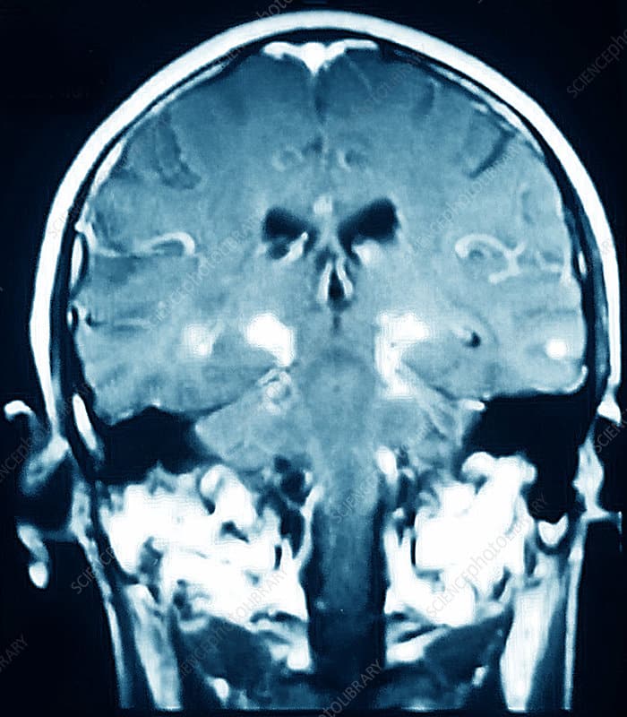
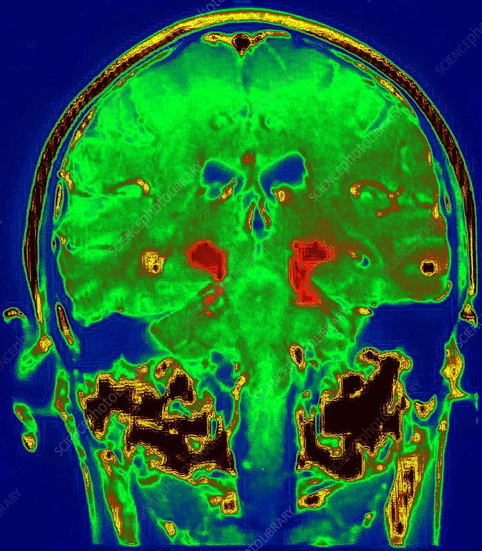
Traditional brain imaging with CT and MRI scans do not show changes in the brain when someone has Parkinsons disease and are generally not helpful in diagnosis. A new kind of brain scan, called a DaT scan, does show changes in persons with Parkinsons disease and may someday become an important tool in diagnosing Parkinsons.
The dopamine transporter, or DaT, scan uses a chemical that labels the dopamine transporter in the area of the brain known as the striatum. Dopamine is a neurochemical that is decreased in persons with Parkinsons disease.
The dopamine transporter, which moves dopamine in and out of cells, is also decreased in the striatum in persons with Parkinsons disease and related disorders. The chemical that labels the transporter is injected into the vein and can be imaged by using something called single photon emission computerized tomography, or SPECT scanning. This technique has been registered in the European Union since 2000 for differentiating a diagnosis of essential tremor and a parkinsonian syndrome. It was approved by the Food and Drug Administration in 2011 for this same indication and recently became available at the OHSU Brain Institute.
Patient And Public Involvement
The OPDC Discovery Cohort is designed by and for patients and is closely linked with the Parkinsons UK local support group. Patient representatives are also involved in the funding/renewal and strategic oversight processes, and sit on the data access panel with casting votes. Results are disseminated to the study participants through annual newsletters, the OPDC website, and series of talks at participants open days.
Also Check: Using Cbd For Parkinsons Disease
How A Diagnosis Is Made
The bedside examination by a neurologist remains the first and most important diagnostic tool for Parkinsons disease . Researchers are working to develop a standard biological marker such as a blood test or an imaging scan that is sensitive and specific for Parkinsons disease.
A neurologist will make the diagnosis based on:
- A detailed history of symptoms, medical problems, current and past medications. Certain medical conditions, as well as some medications, can cause symptoms similar to Parkinsons.
- A detailed neurological examination during which a neurologist will ask you to perform tasks to assess the agility of arms and legs, muscle tone, gait and balance, to see if:
- Expression and speech are animated.
- Tremor can be observed in your extremities at rest or in action.
- There is stiffness in extremities or neck.
- You can maintain your balance and examine your posture.
You May Like: Do Parkinson’s Symptoms Come And Go
I Have Pd And Several Symptoms Should I Get A Datscan
Likely no. There is no need for DaTscan when your history and exam suggest Parkinsons disease and you meet the diagnostic criteria. Occasionally, if signs and symptoms are mild or you dont meet the diagnostic criteria, your doctor will refer you for a DaT scan. Keep in mind that ultimately the diagnosis is based on your history and physical exam. The DaT scan is most commonly used to complete the picture and is not a test for a diagnosis.
What Tests Diagnose Parkinson’s Disease

There currently are no tests that can definitively diagnose Parkinsons Disease. A diagnosis is based on the clinical findings of your physician in combination with your report on the symptoms you are experiencing.
In situations where an older person presents with the typical features of Parkinsons and they are responsive to dopamine replacement therapy, there is unlikely to be any benefit to further investigation or imaging.
Recommended Reading: What State Has Highest Rate Of Parkinson’s
Its Amazing The Difference It Made
Jimmy Hiner struggled for decades with a tremor in his right hand that affected his ability to function. After being diagnosed by experts at the ODonnell Brain Institute, he became the first patient in Texas to undergo a procedure called high-intensity focused ultrasound , which uses MRI-guided ultrasound waves to precisely target an area in the brain linked to Parkinsons disease and essential tremor.
Dont Miss: Does Sam Waterston Have Parkinsons
What Medications And Treatments Are Used
Medication treatments for Parkinsons disease fall into two categories: Direct treatments and symptom treatments. Direct treatments target Parkinsons itself. Symptom treatments only treat certain effects of the disease.
Medications
Medications that treat Parkinsons disease do so in multiple ways. Because of that, drugs that do one or more of the following are most likely:
Several medications treat specific symptoms of Parkinsons disease. Symptoms treated often include the following:
- Erectile and sexual dysfunction.
- Hallucinations and other psychosis symptoms.
Deep brain stimulation
In years past, surgery was an option to intentionally damage and scar a part of your brain that was malfunctioning because of Parkinsons disease. Today, that same effect is possible using deep-brain stimulation, which uses an implanted device to deliver a mild electrical current to those same areas.
The major advantage is that deep-brain stimulation is reversible, while intentional scarring damage is not. This treatment approach is almost always an option in later stages of Parkinsons disease when levodopa therapy becomes less effective, and in people who have tremor that doesnt seem to respond to the usual medications.
Experimental treatments
Researchers are exploring other possible treatments that could help with Parkinsons disease. While these arent widely available, they do offer hope to people with this condition. Some of the experimental treatment approaches include:
You May Like: Animal Models Of Parkinson’s Disease
T Mri Measures Changes In Brains Of Patients With Parkinsons Disease
Researchers at the University of Cambridge used a new ultra-high strength 7T MRI scanner at the Wolfson Brain Imaging Centre to measure changes in the brains of people with Parkinsons disease, progressive supranuclear palsy , or in good health. The study, published in Movement Disorders, suggests that 7T MRI scanners could be used to help identify those patients with Parkinsons disease and similar conditions most likely to benefit from new treatments for previously untreatable symptoms.
Patients with Parkinsons disease and PSP are often treated with drugs such as L-DOPA, which compensate for the severe loss of dopamine. But, dopamine treatment does little for many of the non-motor symptoms. That is why scientists have begun to turn their attention to noradrenaline, a chemical that plays a critical role in brain functions including attention and arousal, thinking and motivation.
Professor James Rowe from the Department of Clinical Neurosciences at the University of Cambridge, who led the study, said, Noradrenaline is very important for brain function. All of our brains supply comes from a tiny region at the back of the brain called the locus coeruleus which means the blue spot. Its a bit like two short sticks of spaghetti half an inch long: its thin, its small, and its tucked away at the very base of the brain in the brain stem.
-
New research indicates that blocking proteins with medications
Mri Brain Scans Detect People With Early Parkinsons
Oxford University researchers have developed a simple and quick MRI technique that offers promise for early diagnosis of Parkinsons disease.
The new MRI approach can detect people who have early-stage Parkinsons disease with 85% accuracy, according to research published in Neurology, the medical journal of the American Academy of Neurology.
At the moment we have no way to predict who is at risk of Parkinsons disease in the vast majority of cases, says Dr Clare Mackay of the Department of Psychiatry at Oxford University, one of the joint lead researchers. We are excited that this MRI technique might prove to be a good marker for the earliest signs of Parkinsons. The results are very promising.
Claire Bale, research communications manager at Parkinsons UK, which funded the work, explains: This new research takes us one step closer to diagnosing Parkinsons at a much earlier stage one of the biggest challenges facing research into the condition. By using a new, simple scanning technique the team at Oxford University have been able to study levels of activity in the brain which may suggest that Parkinsons is present. One person every hour is diagnosed with Parkinsons in the UK, and we hope that the researchers are able to continue to refine their test so that it can one day be part of clinical practice.
We think that our MRI test will be relevant for diagnosis of Parkinsons
Dr Michele Hu
Don’t Miss: Does Parkinson’s Run In The Family
Dark Pigment In Brain Cells
The brain cells affected by Parkinsons contain a pigment called neuromelanin. This pigment gives the cells a characteristic dark colouring.
The technique may be sensitive enough to monitor the progression of Parkinsons.
As these cells are lost in Parkinsons, this pigmentation is reduced. Recent research has suggested MRI may be sensitive enough to detect this change.
However, there are many different machines and methods for taking MRI brain scans, which could affect how accurate the technique is at detecting Parkinsons.
The team developed ways to standardise results from different types of machine to increase the accuracy of the technique as a diagnostic tool.
They also discovered that the technique may be sensitive enough to monitor the progression of Parkinsons.
Read Also: On Off Phenomenon
Innovative Treatment Eliminates Essential Tremor
Rich Shaefers diagnosis of essential tremor, which causes shaking, made daily tasks like eating and getting dressed difficult. An innovative treatment that uses focused ultrasound to penetrate the skull and treat the brain with no incisions allowed him to look toward a future not focused on his tremor.
Dont Miss: Weighted Silverware
Recommended Reading: Early-onset Vs Late-onset Parkinson
Is Parkinsons Diagnosed In The Brain
Parkinsons disease is one of the most challenging neurological disorders to diagnose and treat. If your doctor suspects you have Parkinsons disease, you will usually be referred to a neurologist for further tests. These tests will involve certain movements and exercises to check your symptoms.
A neurologist will look for motor symptoms such as:
- A tremor that occurs at rest
- Slowed movement
- Muscle stiffness
If you have two or more of these symptoms and your doctor has taken blood tests to rule out other causes, its likely you will be diagnosed with Parkinsons disease. Your symptoms will be closely monitored to see any progression of Parkinsons disease, which can take years.
Recommended Reading: Clinical Trials On Parkinsons Disease
Fast Recovery Immediate Success

The procedure takes place under local anaesthetic and you will need a one night hospital stay afterwards. There is a period of about one month afterwards when you may notice some minor issues that gradually improve and we advise you not to drive for one month after the procedure.
The treatment works immediately and the results are expected to be long-lasting.
Also Check: What Tests Are Done For Parkinson’s
Researchers Examine How Parkinsons Disease Alters Brain Activity Over Time
Tracking neural changes could help researchers test therapies that slow disease progression.
Neuroscientists peered into the brains of patients with Parkinsons disease and two similar conditions to see how their neural responses changed over time. The study, funded by the NIHs Parkinsons Disease Biomarkers Program and published in Neurology, may provide a new tool for testing experimental medications aimed at alleviating symptoms and slowing the rate at which the diseases damage the brain.
If you know that in Parkinsons disease the activity in a specific brain region is decreasing over the course of a year, it opens the door to evaluating a therapeutic to see if it can slow that reduction, said senior author David Vaillancourt, Ph.D., a professor in the University of Floridas Department of Applied Physiology and Kinesiology. It provides a marker for evaluating how treatments alter the chronic changes in brain physiology caused by Parkinsons.
For decades, the field has been searching for an effective biomarker for Parkinsons disease, said Debra Babcock, M.D., Ph.D., program director at the NIHs National Institute of Neurological Disorders and Stroke . This study is an example of how brain imaging biomarkers can be used to monitor the progression of Parkinsons disease and other neurological disorders.
The study was supported by the NIH .
NIHTurning Discovery Into Health®
You May Like: How To Get Checked For Parkinsons Disease
What Are The Risk Factors For Parkinsons
There are a few known risk factors for Parkinsons. These include:
- having a family history of Parkinsons
- being over 60 years
- having been exposed to herbicides, pesticides, and other toxins
Its important to note that these risk factors only cause a slight increase in risk. Having one or more risk factors isnt an indicator youll develop Parkinsons. However, if youre concerned about your risk for Parkinsons, talk with a doctor.
Also Check: Does Parkinson’s Cause Delusions
Clinical Mr Imaging In Parkinsons Disease: How Useful Is The Swallow Tail Sign
Institute of Neurogenetics, University of Lübeck, Lübeck, Germany
Department of Neurology, University Medical Center Schleswig-Holstein, Lübeck, Germany
Center for Brain, Behavior, and Metabolism, University of Lübeck, Lübeck, Germany
Institute of Neurogenetics, University of Lübeck, Lübeck, Germany
Department of Neurology, University Medical Center Schleswig-Holstein, Lübeck, Germany
Department of Psychiatry and Psychotherapy, University Medical Center Schleswig-Holstein, Lübeck, Germany
Institute of Neurogenetics, University of Lübeck, Lübeck, Germany
Department of Neurology, University Medical Center Schleswig-Holstein, Lübeck, Germany
Institute of Neurogenetics, University of Lübeck, Lübeck, Germany
Department of Neurology, University Medical Center Schleswig-Holstein, Lübeck, Germany
Institute of Neurogenetics, University of Lübeck, Lübeck, Germany
Department of Neurology, University Medical Center Schleswig-Holstein, Lübeck, Germany
Center for Brain, Behavior, and Metabolism, University of Lübeck, Lübeck, Germany
Department of Neurology, University Medical Center Schleswig-Holstein, Lübeck, Germany
Center for Brain, Behavior, and Metabolism, University of Lübeck, Lübeck, Germany
Institute of Psychology II, University Medical Center Schleswig-Holstein, Lübeck, Germany
Institute of Neurogenetics, University of Lübeck, Lübeck, Germany
Department of Neurology, University Medical Center Schleswig-Holstein, Lübeck, Germany
Correspondence
Correspondence
Conflict Of Interest Statement
EB has equity stake in Motac holding Ltd. and receives consultancy payments from Motac Neuroscience Ltd., companies which pre-clinical activity has no relationship with the present study.
The remaining authors declare that the research was conducted in the absence of any commercial or financial relationships that could be construed as a potential conflict of interest.
Read Also: Can You Live With Parkinson’s
Mri Brain Scans Detect People With Early Parkinson’s
Oxford University researchers have developed a simple and quick MRI technique that offers promise for early diagnosis of Parkinson’s disease.
The new MRI approach can detect people who have early-stage Parkinson’s disease with 85% accuracy, according to research published in Neurology, the medical journal of the American Academy of Neurology.
‘At the moment we have no way to predict who is at risk of Parkinson’s disease in the vast majority of cases,’ says Dr Clare Mackay of the Department of Psychiatry at Oxford University, one of the joint lead researchers. ‘We are excited that this MRI technique might prove to be a good marker for the earliest signs of Parkinson’s. The results are very promising.’
Claire Bale, research communications manager at Parkinson’s UK, which funded the work, explains: ‘This new research takes us one step closer to diagnosing Parkinson’s at a much earlier stage one of the biggest challenges facing research into the condition. By using a new, simple scanning technique the team at Oxford University have been able to study levels of activity in the brain which may suggest that Parkinson’s is present. One person every hour is diagnosed with Parkinson’s in the UK, and we hope that the researchers are able to continue to refine their test so that it can one day be part of clinical practice.’
We think that our MRI test will be relevant for diagnosis of Parkinson’s
Dr Michele Hu
Brain Mr Contribution To The Differential Diagnosis Of Parkinsonian Syndromes: An Update
Giovanni Rizzo
1IRCCS Istituto delle Scienze Neurologiche, Bellaria Hospital, Bologna, Italy
2Neurology Unit, Department of Biomedical and Neuromotor Sciences, University of Bologna, Bologna, Italy
3Functional MR Unit, Policlinico S.Orsola-Malpighi, Bologna, Italy
4Department of Biomedical and Neuromotor Sciences, University of Bologna, Bologna, Italy
5Department of Diagnostic Imaging, Pia Fondazione di Culto e Religione Card. G. Panico, Tricase, Italy
6Department of Neurology, Kings College NHS Foundation Trust, London, UK
7Department of Neurology, Queen Elizabeth Hospital, Lewisham and Greenwich NHS Trust, London, UK
8Department of Neurology and Health Science Research, Mayo Clinic, Rochester, MN, USA
9Department of Clinical Research in Neurology, University of Bari, Pia Fondazione di Culto e Religione Card. G. Panico, Tricase, Italy
10Department of Basic Medical Science, Neuroscience and Sense Organs, University of Bari, Bari, Italy
Abstract
1. Introduction
Many putative biomarkers derived from genetic-epigenetic, neurophysiological, and imaging techniques have been evaluated in order to determine their diagnostic accuracy in discriminating PD from APSs . Brain magnetic resonance represents one of the best putative sources of biomarkers in this field, because of its relative feasibility, the absence of invasiveness, and the availability in different clinical settings.
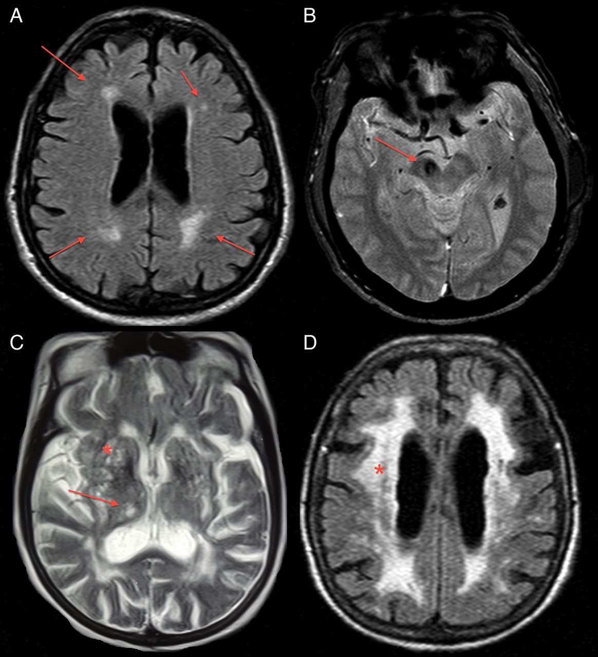
2. Qualitative Brain MRI
Also Check: What Is The Pathophysiology Of Parkinson’s Disease
Preparing For A Parkinsons Mri
A Parkinsons MRI is completely painless, but you do have to lie still while being scanned. Some patients feel claustrophobic in this situation. If youre worried about that, talk with your doctor about the possibility of having an anti-anxiety medication before the procedure.
On the day of the appointment, follow any instructions provided to you by your doctor. Remove metal jewelry and dont wear make-up as that can also have metal in it. If you are in the advanced stages of Parkinsons or if you are taking a sedative, you should arrange transportation to and from the appointment.
What Is Focused Ultrasound

Focused ultrasound is a non-invasive, image-guided surgical technology that harnesses the power of ultrasound energy. Using a specially designed helmet, ultrasound is delivered across the skull to deep brain targets without requiring scalpels or incisions. At Sunnybrook focused ultrasound is delivered in an MRI so real time image guidance can be used.
Recommended Reading: Is Restless Legs A Symptom Of Parkinson’s
